


Thrombocytopenia Panel - Expanded
Summary and Pricing 
Test Method
Exome Sequencing with CNV Detection| Test Code | Test Copy Genes | Panel CPT Code | Gene CPT Codes Copy CPT Code | Base Price | |
|---|---|---|---|---|---|
| 10393 | Genes x (33) | 81479 | 81404(x1), 81406(x2), 81479(x63) | $990 | Order Options and Pricing |
Pricing Comments
We are happy to accommodate requests for testing single genes in this panel or a subset of these genes. The price will remain the list price. If desired, free reflex testing to remaining genes on panel is available. Alternatively, a single gene or subset of genes can also be ordered via our Custom Panel tool.
An additional 25% charge will be applied to STAT orders. STAT orders are prioritized throughout the testing process.
Click here for costs to reflex to whole PGxome (if original test is on PGxome Sequencing platform).
Click here for costs to reflex to whole PGnome (if original test is on PGnome Sequencing platform).
Turnaround Time
3 weeks on average for standard orders or 2 weeks on average for STAT orders.
Please note: Once the testing process begins, an Estimated Report Date (ERD) range will be displayed in the portal. This is the most accurate prediction of when your report will be complete and may differ from the average TAT published on our website. About 85% of our tests will be reported within or before the ERD range. We will notify you of significant delays or holds which will impact the ERD. Learn more about turnaround times here.
Targeted Testing
For ordering sequencing of targeted known variants, go to our Targeted Variants page.
Clinical Features and Genetics
Clinical Features
Inherited thrombocytopenias (IT) comprise a heterogeneous group of disorders characterized by platelet counts below the lower limit of normal, that's below 150,000/µL (150 x 109/L) in adults. Bleeding manifestations of thrombocytopenia range from mild to severe and may include excessive bruising (purpura), petechiae, prolonged bleeding from cuts or from surgical procedures, spontaneous nose bleeds, and in women, heavy menstrual flows. About half of ITs are syndromic disorders characterized by other physical and neurological anomalies, or immunodeficiencies (Balduini et al. 2013. PubMed ID: 23397552). Over 30 genes are known to be associated with ITs, and pathogenic variants in known genes are found in only about 50% of cases (Kunishima and Saito. 2006. PubMed ID: 16169642; Noris and Pecci 2017. PubMed ID: 29222283). Noris and Pecci divide ITs into three groups: forms characterized by only platelet deficiencies, syndromic ITs with additional congenital defects, and ITs associated with increased risk of developing additional disease such as myelodysplastic syndrome (MDS) and acute leukemia (AL). For additional information regarding inherited hematologic malignancies, see Churpek et al. 2013. PubMed ID: 22691122; Furutani and Shimamura. 2017. PubMed ID: 28297620. It is important to distinguish ITs from immune/idiopathic thrombocytopenias (ITP) in order to inform clinical management and identify potential at risk family members.
Genetics
The list of genes associated with inherited thrombocytopenias continues to grow. This panel is similar to our Thrombocytopenia Panel, but includes additional genes that are associated with disorders having clinical features other than thrombocytopenia, and genes with variants causative for thrombocytopenia, but that are found in relatively few patients. The genes included in this panel have been associated with both syndromic and non-syndromic forms of inherited thrombocytopenia and represent the best documented forms of inherited thrombocytopenia reported in the literature. Thrombocytopenias are typically divided into three distinct groups based upon platelet size: large/macrothrombocytopenias, small/microthrombocytopenias, and thrombocytopenias with normal sized platelets.
The majority of pathogenic variants reported in the thrombocytopenia genes in this panel are missense and nonsense variants; large deletions and duplications are rare. Some exceptions are described below.
Macrothrombocytopenias
ACTN1 — α actinin-related thrombocytopenias are inherited in an autosomal dominant manner and are characterized by pathogenic variants in the ACTN1 gene. These variants alter actin filament assembly and platelet cytoskeletal structure causing macrothrombocytopenia (Kunishima et al. 2013. PubMed ID: 23434115). Patients with α actinin-related thrombocytopenias display mild bleeding and macrothrombocytopenia, but have no other distinguishing traits thereby making genetic diagnosis necessary for these individuals.
ABCG5, ABCG8 — Sitosterolemia is caused by pathogenic variants in either the ABCG5 or ABCG8 genes, and is characterized by defects in the ABC carrier protein that is important for sterol metabolism. Hemolytic anemia, stomatocyte formation, and macrothrombocytopenia are common hematologic findings and may be the only clinically observed symptoms in patients (Wang et al. 2014. PubMed ID:24166850; Escolá-Gil et al. 2014. PubMed ID:24821603). Inheritance is autosomal recessive.
CD36 – The CD36 gene encodes platelet surface-expressed glycoprotein IV (GPIV) which serves as the primary receptor for platelet – collagen interactions (Tandon et al. 1989. PubMed ID: 2468670). Pathogenic variants in CD36 are associated with autosomal recessive Platelet Glycoprotein IV Deficiency that is characterized by macrothrombocytopenia and variable bleeding tendencies (Yufu et al. 1990. PubMed ID: 2316511). Pathogenic variants in CD36 comprise missense, nonsense, and both small and large deletions that result in reduced GPIV production and/or surface expression. Pathogenic variants in CD36 are found at a high rate in African populations; these variants are also associated with susceptibility and severity of malaria (Aitman et al. 2000. PubMed ID: 10890433).
FLI1 — Deletions encompassing chromosome 11q are associated with Paris-Trousseau syndrome and Jacobsen syndrome- overlapping disorders characterized by thrombocytopenia, facial and cardiac dysmorphism, growth restrictions and intellectual disability (Krishnamurti et al. 2001. PubMed ID:11279643; Favier et al. 2003. PubMed ID:14597985). Platelets in patients with Paris-Trousseau syndrome and Jacobsen syndrome are large and contain giant alpha granules. The FLI1 gene encodes a transcription factor involved in megakaryocyte differentiation. FLI1 resides on chromosome 11q and is located in the deleted region found in Paris-Trousseau syndrome and Jacobsen syndrome patients. Hemizygous loss of FLI1 and monoallelic expression are thought to contribute to the platelet phenotype found in Paris-Trousseau syndrome and Jacobsen syndrome patients (Raslova et al. 2004. PubMed ID:15232614). Missense variants in FLI1 have also been reported in families with bleeding phenotypes and platelet dense granule secretion defects (Stockley et al. 2013. PubMed ID:24100448). Inheritance is autosomal dominant, though a case of recessive inheritance in a consanguineous family was reported (Stevenson et al. 2015. PubMed ID:26316623).
FLNA — Pathogenic variants in FLNA are associated with periventricular nodular heterotopia (PVNH) and otopalatodigital spectrum disorders. The FLNA protein, Filamin A, cross-links actin filaments in the cytoskeleton. Patients with filaminopathies A are characterized by skeletal dysplasia, congenital malformations and mental retardation in some cases (Robertson et al. 2003. PubMed ID:12612583; Kyndt et al. 2007. PubMed ID:17190868). These patients may also have macrothrombocytopenia and abnormal platelets with a rounded morphology, heterozygous α-granules, and occasional giant granules (Nurden et al. 2011. PubMed ID:21960593). These abnormalities may be caused by abnormal FLNA protein disrupting normal megakaryocyte differentiation (Nurden et al. 2011. PubMed ID:21960593). Inheritance is X-linked dominant.
GFI1B — The GFI1B protein is a transcription factor essential for erythropoiesis and megakaryopoiesis. Pathogenic variants in GFI1B are associated with abnormal megakaryocytes and platelets. These variants result in macrothrombocytopenia with a variable bleeding phenotype and the presence of red cell anisopoikilocytosis (Stevenson et al. 2013. PubMed ID:23927492; Monteferrario et al. 2014. 24325358). Inheritance is autosomal dominant. Abnormal GFI1B protein may act in a dominant negative manner resulting in thrombocytopenia with alpha-granule deficiency and the characteristic gray appearance found in Gray Platelet syndrome (Stevenson et al. 2013. PubMed ID:23927492; Monteferrario et al. 2014. PubMed ID:24325358).
GNE — Variants in GNE are typically associated with recessive GNE myopathy that presents as a slowly progressive distal weakness in young adults in their second or third decades of life (Mori-Yoshimura et al. 2014. PubMed ID: 24656604). However, some patients with GNE myopathy were reported to have mild, asymptomatic thrombocytopenia (Mori-Yoshimura et al. 2014. PubMed ID: 25303967). Two recent studies report patients with severe congenital thrombocytopenia with no clinical evidence of GNE myopathy who harbored homozygous variants in GNE (Revel-Vilk et al. 2018. PubMed ID: 30171045; Futterer et al. 2018. PubMed ID: 29941673). In addition to large platelets, patients showed bruising and bleeding tendencies though platelet aggregation appeared to be unaffected. GNE is expressed in all hematopoietic cells and encodes the bi-functional enzyme UDP-N-acetylglucosamine 2-epimerase that is important for sialic acid biosynthesis which is required for normal sialylation in hematopoietic cells. Dysregulated sialic acid biosynthesis has been associated with thrombocytopenia (Li et al. 2017. PubMed ID: 28494777) and may be the mechanism of GNE-related thrombocytopenia.
MYH9 — May-Hegglin Anomaly, Sebastian Syndrome, Fechtner Syndrome, and Epstein Syndrome. Inheritance is autosomal dominant. Clinical features may include high tone deafness, cataracts, leukocyte inclusions, and kidney disease leading in some cases to renal failure. Pathogenic variants affecting the head domain of the Myosin-IIA protein are associated with a higher risk of nephropathy and deafness than variants affecting the tail domain (Pecci et al. 2014. 24186861).
GP1BA, GP1BB, GP9 — Bernard Soulier Syndrome and Platelet Type von Willebrand Disease (PT-VWD, GP1BA), and Giant Platelet Syndrome. Inheritance is generally autosomal recessive. Pathogenic variants in GP1BA are also associated with PT-VWD and are inherited in an autosomal dominant manner.
GATA1 — X-linked thrombocytopenia. Inheritance is X-linked recessive. Clinical features include erythrocytic anemia, globin gene transcription defects, and porphyria.
ITGA2B, ITGB3 — Glanzmann thrombasthenia (GT) is inherited in an autosomal recessive manner and is caused by pathogenic variants in the genes encoding the αIIb and β3 integrins that result in failure of platelets to aggregate. Platelets in GT patients generally have normal morphology and are present at normal levels, however in rare cases, variants in ITGA2B and ITGB3 result in dominant macrothrombocytopenia without a severe GT phenotype (Nurden et al. 2013. PubMed ID: 23929305).
NBEAL2 — Gray Platelet Syndrome (GPS) is characterized primarily by mild to severe bleeding tendency, and moderate thrombocytopenia with enlarged platelets that lack α-granules (Raccuglia. 1971. PubMed ID:5129551; Breton-Gorius et al. 1981. PubMed ID:7468753; Gunay-Aygun et al. 2010. PubMed ID:20709904). Platelets appear gray in color in peripheral blood smears which is attributed to a lack of platelet granules. Inheritance is autosomal recessive.
PRKACG — Bleeding Disorder, Platelet-Type, 19 (BDPLT19). The PRKACG gene encodes the γ-catalytic subunit of the cyclic adenosine monophosphate-dependent protein kinase. A missense variant in PRKACG, c.222C>G (p.Ile74Met), was reported in one family with severe macrothrombocytopenia and early childhood onset (Manchev et al. 2014. PubMed ID:25061177). Platelet formation was altered in these patients suggesting that the PRKACG protein plays a role in platelet biogenesis (Manchev et al. 2014. PubMed ID:25061177). Inheritance is autosomal recessive and to date, no other variants in PRKACG have been reported to be causative for thrombocytopenia.
TUBB1 — TUBB1 encodes the megakaryocyte restricted β1 tubulin which forms heterodimers with a tubulin in microtubules that help maintain platelet morphology (Italiano et al. 2003. PubMed ID:12586623). Pathogenic variants in TUBB1 have been reported in a small number of patients with macrothrombocytopenia, but who have no other distinguishing traits thereby making genetic diagnosis necessary for these patients (Kunishima et al. 2009. PubMed ID:18849486). Inheritance is autosomal dominant.
Microthrombocytopenias
FYB1 — Pathogenic variants in FYB1 are associated with autosomal recessive Thrombocytopenia 3. To date, one nonsense variant and one small deletion resulting in premature protein truncation have been reported, each in different consanguineous families with small platelet thrombocytopenia (Levin et al. 2013. PubMed ID: 23650215; Hamamy et al. 2014. PubMed ID: 25516138). Bleeding tendency varies among patients and may manifest as heavy menstrual bleeding in some females.
PTPRJ—Two siblings with microthrombocytopenia were recently reported to have compound heterozygous loss of function variants in the PTPRJ gene (Marconi et al. 2019. PubMed ID: 30591527). Disease presented as nonsyndromic thrombocytopenia with mild to moderate bleeding tendencies, small platelets, and impaired megakaryocyte maturation and platelet formation. The patients showed a severe depletion of PTPRJ protein in blood cells, and PTPRJ protein levels in the carrier parents were decreased by ~ 50%. To date, these are the only patients reported to have PTPRJ-related thrombocytopenia. PTPRJ is a receptor-type protein tyrosine phosphatase expressed in several different cell types including hematopoietic cells that acts as a master regulator of Src family kinases in platelets and megakaryocytes (Senis. 2013. PubMed ID: 24015866).
WAS — Wiskott-Aldrich syndrome, X-linked thrombocytopenia. Inheritance is X-linked recessive. Clinical features may include eczema, recurrent bacterial and viral infections, severe hemorrhaging, autoimmune disease such as hemolytic anemia or immune thrombocytopenic purpura, lymphomas, and X-linked Congenital Neutropenia.
Thrombocytopenias with Normal Platelet Size
CYCS — Cytochrome-C gene related thrombocytopenia. Inheritance is autosomal dominant.
ETV6 — Pathogenic variants in ETV6 have been identified in several families with autosomal dominant thrombocytopenia (Thrombocytopenia 5) who have mild-to-moderate bleeding tendencies and an increased risk of developing hematologic malignancies including MDS, Acute Myeloid Leukemia (AML) and acute lymphoblastic leukemia (ALL) (Zhang et al. 2015. PubMed ID: 25239263; Topka et al. 2015. PubMed ID: 26102509; Noetzli et al. 2015. PubMed ID: 25807284). Thrombocytopenia is usually present in early childhood while malignancies may develop throughout life.
ANKRD26, MASTL — Thrombocytopenia 2 (THC2) is characterized by moderately low platelet counts (family averages = 40-60/nl) (Savoia et al. 1999. PubMed ID:10521306; Drachman et al. 2000. PubMed ID:10891439). Platelets are of normal size. Patients often bruise easily and have moderate bleeding problems. Thrombopoietin levels are mildly elevated. Pathogenic variants in ANKRD26 are associated with predisposition to MDS and AML; up to a 30 fold increase in the frequency of MDS and AML has been reported in patients with ANKRD26 gene pathogenic variants (Noris et al. 2011. PubMed ID:21467542; Noris et al. 2013. PubMed ID:24030261; Marquez et al. 2014. PubMed ID:24628296). Pathogenic variants in the MASTL gene cause autosomal dominant thrombocytopenia (Gandhi et al. 2003. PubMed ID: 12890928). To date, one causative missense variant (Asp167Glu) has been reported in humans.
RUNX1 — Familial Thrombocytopenia with Predisposition to AML. Inheritance is autosomal dominant. Over 40% of patients with germline pathogenic variants in the RUNX1 gene develop MDS/AML at a mean age of ~ 33 years (Churpek et al. 2013. PubMed ID:22691122).
MECOM — Pathogenic variants in MECOM are associated with autosomal dominant radioulnar synostosis with amegakaryocytic thrombocytopenia 2 (RUSAT2) an inherited form of bone marrow failure (Niihori et al. 2015. PubMed ID: 26581901). MECOM encodes the transcription factor EVI1 which plays a key role in hematopoiesis (Katoaoka and Kurokawa. 2012. PubMed ID: 22494115). To date, 3 different missense variants in the MECOM gene that affect the 8th zinc finger domain have been identified in different families with RUSAT2; functional studies of the missense variants demonstrated DNA-binding deficiencies and altered reporter gene expression compared to wildtype (Niihori et al. 2015. PubMed ID: 26581901). RUSAT2 is also associated with severe anemia and hearing abnormalities in some cases (Niihori et al. 2015. PubMed ID: 26581901).
MPL — Congenital Amegakaryocytic Thrombocytopenia (CAMT). Inheritance is autosomal recessive. Pathogenic variants in MPL have been identified in ~ 60% of patients. Clinical features include absent or reduced megakaryocytes and development of aplastic anemia and pancytopenia. AML has been reported in some patients (Ballmaier and Gerheshausen. 2009. PubMed ID:19388932).
ADAMTS13 — Thrombotic Thrombocytopenic Purpura (TTP), often described as Upshaw-Schulman syndrome (USS), is a rare blood condition characterized by frequent relapses of fever, platelet thrombi in microvasculature, hemolytic anemia, consumptive thrombocytopenia, neurologic symptoms, renal disease, and occasionally organ failure (Levy et al. 2001. PubMed ID:11586351). Inheritance is autosomal recessive.
HOXA11 — Congenital Thrombocytopenia with Radioulnar Synostosis (CTRUS). HOXA11 is a member of the homeobox family of regulatory proteins that are essential for bone formation and hematopoiesis. A variant in the HOXA11 gene, c.872delA (p.Asn291Thrfs*4), was found in two unrelated families with radioulnar synostosis and amegakaryocytic thrombocytopenia (Thompson and Nguyen. 2000. PubMed ID:11101832). To date, no other variants in the HOXA11 gene have been identified in patients with CTRUS, and it is likely that other genes are also associated with the CTRUS phenotype. Inheritance is autosomal dominant.
RBM8A — Thrombocytopenia with Absent Radius (TAR) (CTRUS). TAR is characterized by thrombocytopenia and absence of the radius with preservation of the thumb. The severity of bleeding episodes tends to diminish in frequency and severity over time. Skeletal abnormalities may extend to the absence of upper limbs and to the hips and knees (Greenhalgh et al. 2002. PubMed ID:12471199). Inheritance is autosomal recessive. Patients with TAR have a compound inheritance including a null allele and a low frequency variant in the RBM8A gene. Reported null alleles comprise deletions of chromosome 1q21.1, which includes the RBM8A gene and is found in most patients (Klopocki et al. 2007. PubMed ID:17236129; Papoulidis et al. 2014. PubMed ID:24220582), a small insertion (c.207_208insAGCG) resulting in premature protein termination (p.Val70Serfs*3) (Albers et al. 2012. PubMed ID: 22366785), and a nonsense variant c.487C>T (p.Arg163*) (Albers et al. 2012. PubMed ID: 22366785). Paring one of these null alleles with one of two noncoding variants on the other allele, either c.-21G>A or c.67+32G>C, is strongly associated with disease (Albers et al. 2012. PubMed ID:22366785). This test includes sequencing of the coding regions of the RBM8A gene and sequencing for the c.-21G>A and c.67+32G>C variants.
Large deletions account for ~24% of the reported RUNX1 gene variants, but in general, large, multi-exon and whole gene deletions are rare among the thrombocytopenia panel genes. In addition to the RUNX1 gene, a few large deletions have also been reported in the GP1BB, MYH9, and WAS genes (Human Gene Mutation Database), but represent a small fraction of the total pathogenic variants reported for these genes.
See individual gene test descriptions for more information on molecular biology of gene products and mutation spectra.
Clinical Sensitivity - Sequencing with CNV PGxome
This test is designed to identify pathogenic variants in the most common genes associated with thrombocytopenias. Due to the array of syndromic and non-syndromic forms of inherited thrombocytopenia, and given a continually growing list of genes found in small numbers of families with thrombocytopenia, the overall clinical sensitivity of this panel is difficult to determine. However, in a recent study of 272 patients with macrothrombocytopenia, ~48% of patients harbored pathogenic variants in either the MYH9 gene (~ 38%), the Bernard Soulier genes (~ 10%), or the GATA1 gene (< 1%) (Kunishima, Thrombocytopenia, ISTH Webinar 2015: WEB150325). In general, ~ 50% of inherited thrombocytopenias are not yet characterized (Balduini et al. 2013. 23397552).
Testing Strategy
This is a Next Generation Sequencing Test. See the Test Method link for detailed lab methodology.
This panel typically provides 98.8% coverage of all coding exons of the genes plus 10 bases of flanking noncoding DNA in all available transcripts along with other non-coding regions in which pathogenic variants have been identified at PreventionGenetics or reported elsewhere. We define coverage as ≥20X NGS reads or Sanger sequencing. PGnome panels typically provide slightly increased coverage over the PGxome equivalent. PGnome sequencing panels have the added benefit of additional analysis and reporting of deep intronic regions (where applicable).
In addition to the regions described above, this testing includes coverage of the following variants that reside in untranslated or deep intronic regions: RBM8A c.-21G>A and c.67+32G>C.
Dependent on the sequencing backbone selected for this testing, discounted reflex testing to any other similar backbone-based test is available (i.e., PGxome panel to whole PGxome; PGnome panel to whole PGnome).
Indications for Test
Patients with a family history of thrombocytopenia who may or may not have other symptoms including immunodeficiencies, and physical or neurological anomalies. Patients with a family history of MDS/AML. This thrombocytopenia panel may also be helpful for patients with thrombocytopenia, but without any family history.
Patients with a family history of thrombocytopenia who may or may not have other symptoms including immunodeficiencies, and physical or neurological anomalies. Patients with a family history of MDS/AML. This thrombocytopenia panel may also be helpful for patients with thrombocytopenia, but without any family history.
Genes
| Official Gene Symbol | OMIM ID |
|---|---|
| ABCG5 | 605459 |
| ABCG8 | 605460 |
| ACTN1 | 102575 |
| ADAMTS13 | 604134 |
| ANKRD26 | 610855 |
| ARPC1B | 604223 |
| CD36 | 173510 |
| CYCS | 123970 |
| ETV6 | 600618 |
| FLI1 | 193067 |
| FLNA | 300017 |
| FYB1 | 602731 |
| GATA1 | 305371 |
| GFI1B | 604383 |
| GNE | 603824 |
| GP1BA | 606672 |
| GP1BB | 138720 |
| GP9 | 173515 |
| HOXA11 | 142958 |
| ITGA2B | 607759 |
| ITGB3 | 173470 |
| MASTL | 608221 |
| MECOM | 165215 |
| MPL | 159530 |
| MYH9 | 160775 |
| NBEAL2 | 614169 |
| PRKACG | 176893 |
| PTPRJ | 600925 |
| RBM8A | 605313 |
| RUNX1 | 151385 |
| SMPD1 | 607608 |
| TUBB1 | 612901 |
| WAS | 300392 |
| Inheritance | Abbreviation |
|---|---|
| Autosomal Dominant | AD |
| Autosomal Recessive | AR |
| X-Linked | XL |
| Mitochondrial | MT |
Diseases
Related Test
| Name |
|---|
| PGxome® |
| Thrombocytopenia Panel |
Citations
- Aitman et al. 2000. PubMed ID: 10890433
- Albers et al. 2012. PubMed ID: 22366785
- Balduini et al. 2013. PubMed ID: 23397552
- Ballmaier and Germeshausen. 2009. PubMed ID: 19388932
- Breton-Gorius et al. 1981. PubMed ID: 7468753
- Churpek et al. 2013. PubMed ID: 22691122
- Drachman et al. 2000. PubMed ID: 10891439
- Escolà-Gil et al. 2014. PubMed ID: 24821603
- Favier et al. 2003. PubMed ID: 14597985
- Furutani and Shimamura. 2017. PubMed ID: 28297620
- Futterer et al. 2018. PubMed ID: 29941673
- Gandhi et al. 2003. PubMed ID: 12890928
- Greenhalgh et al. 2002. PubMed ID: 12471199
- Gunay-Aygun et al. 2010. PubMed ID: 20709904
- Hamamy et al. 2014. PubMed ID: 25516138
- Human Gene Mutation Database (Bio-base).
- Italiano et al. 2003. PubMed ID: 12586623
- Kataoka and Kurokawa. 2012. PubMed ID: 22494115
- Klopocki et al. 2007. PubMed ID: 17236129
- Krishnamurti et al. 2001. PubMed ID: 11279643
- Kunishima and Saito. 2006. PubMed ID: 16169642
- Kunishima et al. 2009. PubMed ID: 18849486
- Kunishima et al. 2013. PubMed ID: 23434115
- Kyndt et al. 2007. PubMed ID: 17190868
- Levin et al. 2013. PubMed ID: 23650215
- Levy et al. 2001. PubMed ID: 11586351
- Li et al. 2017. PubMed ID: 28494777
- Manchev et al. 2014. PubMed ID: 25061177
- Marconi et al. 2019. PubMed ID: 30591527
- Marquez et al. 2014. PubMed ID: 24628296
- Monteferrario et al. 2014. PubMed ID: 24325358
- Mori-Yoshimura et al. 2014. PubMed ID: 25303967
- Mori-Yoshimura et al. 2014. PubMed ID: 24656604
- Niihori et al. 2015. PubMed ID: 26581901
- Noetzli et al. 2015. PubMed ID: 25807284
- Noris and Pecci. 2017. PubMed ID: 29222283
- Noris et al. 2011. PubMed ID: 21467542
- Noris et al. 2013. PubMed ID: 24030261
- Nurden et al. 2011. PubMed ID: 21960593
- Nurden et al. 2013. PubMed ID: 23929305
- Papoulidis et al. 2014. PubMed ID: 24220582
- Pecci et al. 2014. PubMed ID: 24186861
- Raccuglia. 1971. PubMed ID: 5129551
- Raslova et al. 2004. PubMed ID: 15232614
- Revel-Vilk et al. 2018. PubMed ID: 30171045
- Robertson et al. 2003. PubMed ID: 12612583
- Savoia et al. 1999. PubMed ID: 10521306
- Senis. 2013. PubMed ID: 24015866
- Stevenson et al. 2013. PubMed ID: 23927492
- Stevenson et al. 2015. PubMed ID: 26316623
- Stockley et al. 2013. PubMed ID: 24100448
- Tandon et al. 1989. PubMed ID: 2468670
- Thompson and Nguyen. 2000. PubMed ID: 11101832
- Topka et al. 2015. PubMed ID: 26102509
- Wang et al. 2014. PubMed ID: 24166850
- Yufu et al. 1990. PubMed ID: 2316511
- Zhang et al. 2015. PubMed ID: 25239263
Ordering/Specimens
Ordering Options
We offer several options when ordering sequencing tests. For more information on these options, see our Ordering Instructions page. To view available options, click on the Order Options button within the test description.
myPrevent - Online Ordering
- The test can be added to your online orders in the Summary and Pricing section.
- Once the test has been added log in to myPrevent to fill out an online requisition form.
- PGnome sequencing panels can be ordered via the myPrevent portal only at this time.
Requisition Form
- A completed requisition form must accompany all specimens.
- Billing information along with specimen and shipping instructions are within the requisition form.
- All testing must be ordered by a qualified healthcare provider.
For Requisition Forms, visit our Forms page
If ordering a Duo or Trio test, the proband and all comparator samples are required to initiate testing. If we do not receive all required samples for the test ordered within 21 days, we will convert the order to the most effective testing strategy with the samples available. Prior authorization and/or billing in place may be impacted by a change in test code.
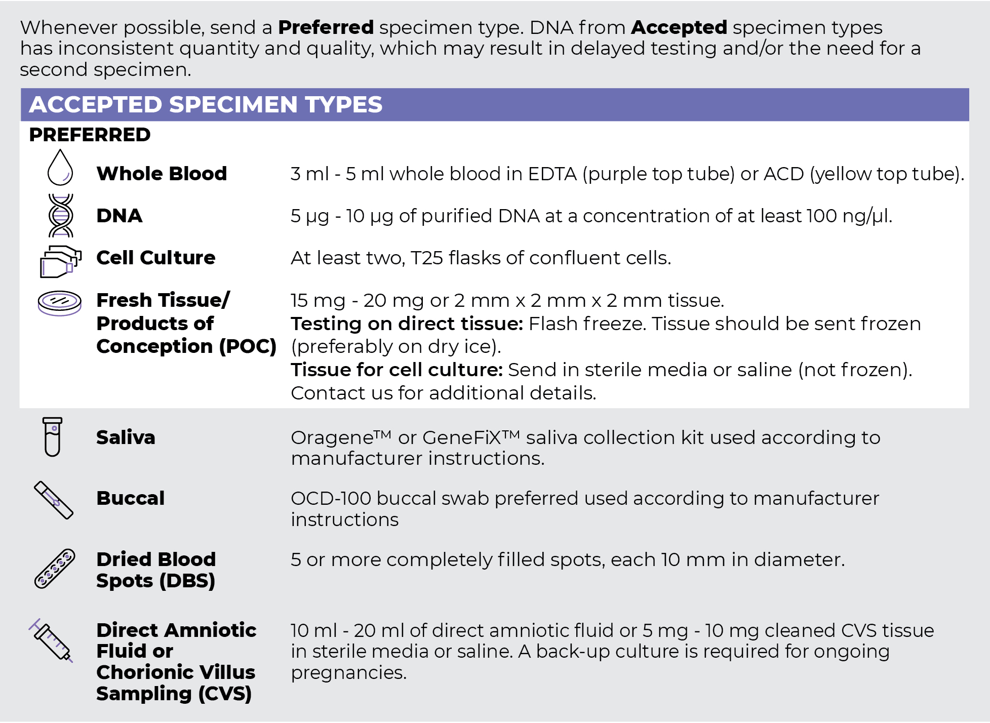
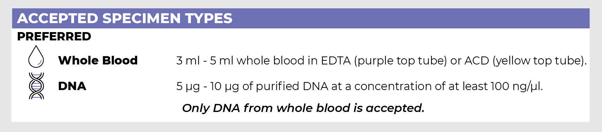
Specimen Types
Specimen Requirements and Shipping Details
PGxome (Exome) Sequencing Panel
PGnome (Genome) Sequencing Panel
ORDER OPTIONS
View Ordering Instructions1) Select Test Type
2) Select Additional Test Options
No Additional Test Options are available for this test.