


Polycystic Liver Disease (PLD) Panel
Summary and Pricing 
Test Method
Exome Sequencing with CNV Detection| Test Code | Test Copy Genes | Panel CPT Code | Gene CPT Codes Copy CPT Code | Base Price | |
|---|---|---|---|---|---|
| 10227 | Genes x (8) | 81479 | 81406(x2), 81407(x1), 81479(x13) | $990 | Order Options and Pricing |
Pricing Comments
We are happy to accommodate requests for testing single genes in this panel or a subset of these genes. The price will remain the list price. If desired, free reflex testing to remaining genes on panel is available. Alternatively, a single gene or subset of genes can also be ordered via our Custom Panel tool.
An additional 25% charge will be applied to STAT orders. STAT orders are prioritized throughout the testing process.
Click here for costs to reflex to whole PGxome (if original test is on PGxome Sequencing platform).
Click here for costs to reflex to whole PGnome (if original test is on PGnome Sequencing platform).
Turnaround Time
3 weeks on average for standard orders or 2 weeks on average for STAT orders.
Please note: Once the testing process begins, an Estimated Report Date (ERD) range will be displayed in the portal. This is the most accurate prediction of when your report will be complete and may differ from the average TAT published on our website. About 85% of our tests will be reported within or before the ERD range. We will notify you of significant delays or holds which will impact the ERD. Learn more about turnaround times here.
Targeted Testing
For ordering sequencing of targeted known variants, go to our Targeted Variants page.
Clinical Features and Genetics
Clinical Features
Isolated polycystic liver disease (PCLD) is one of the three clinical entities of polycystic liver disease, a collection of disorders characterized by development of multiple hepatic cysts in adulthood due to embryonic ductal plate malformation of the intrahepatic biliary tree (van Keimpema et al. 2011. PubMed ID: 20408955; Cnossen et al. 2014. PubMed ID: 24886261). A polycystic liver can be also found in Von Meyenburg complexes (VMC; also termed microhamartomas) characterized by small, nonhereditary nodular cystic lesions; and autosomal dominant polycystic kidney disease (ADPKD) with concurrent hepatic cysts in over 80% of cases as the most frequent extra-renal manifestation.
The presentation of isolated polycystic liver disease (PCLD) is restricted to the liver in the absence of polycystic kidneys. In contrast, ADPKD is a multi-systemic disorder with polycystic kidneys as the major feature. Polycystic liver disease is the major extra-renal phenotype in ADPKD. Therefore, this panel includes PCLD and ADPKD genes (Cornec-Le Gall et al. 2019. PubMed ID: 30819518).
Genetics
Isolated polycystic liver disease (PCLD) is an autosomal dominant disorder caused by pathogenic variants in PRKCSH, SEC63, ALG8 or LRP5 (Cnossen et al. 2014. PubMed ID: 24886261; Cnossen et al. 2014. PubMed ID: 24706814; Besse et al. 2017. PubMed ID: 28375157).
PRKCSH (16 coding exons) and SEC63 (21 coding exons) encode the beta-subunit of glucosidase II (a protein kinase C substrate) and Sec63p, respectively. Both proteins are located within the endoplasmic reticulum (ER) and are responsible for quality control and translocation of glycoproteins into the ER. Pathogenic variants in PRKCSH and SEC63 account for about 35% of PCLD patients (Besse et al. 2017. PubMed ID: 28375157).
The ALG8 gene (13 coding exons) encodes α-1,3-glucosyltransferase. Similarly to PRKCSH and SEC63, ALG8 (asparagine-linked glycosylation 8) is an ER integral membrane protein in the protein biogenesis pathway. To date, documented genetic defects in PRKCSH, SEC63, and ALG8 include truncating changes (nonsense and frame-shifting small deletion/insertions) and missense substitutions. No large deletions or duplications have been reported in these three genes yet.
The LRP5 gene (23 coding exons) encodes a transmembrane low-density lipoprotein receptor, which plays a key role in skeletal homeostasis. Many bone density related diseases such as autosomal dominant osteopetrosis are caused by pathogenic variants in this gene. LRP5 defects also cause hepatic cystogenesis (clinically diagnosed as PCLD) due to deregulation of the canonical wingless signaling pathway (Cnossen et al. 2014. PubMed ID: 24706814). So far only missense LRP5 variants have been reported in PCLD.
Pathogenic variants in the PKD1 (46 coding exons) and PKD2 (15 coding exons) gene cause approximately 90% of ADPKD (Rossetti et al. 2007. PubMed ID: 17582161; Audrézet et al. 2012. PubMed ID: 22508176). Both genes encode members of the polycystin protein family, which together play an important role in renal tubular development. These pathogenic variants have been found across the whole coding region of both genes. Truncated variants (nonsense, typical splicing and frame-shifting small deletion/insertions) are the majority of PKD1 and PKD2 defects although missense and small in-frame changes are also commonly found. Large deletions and duplications have been reported, but are relatively uncommon (Ariyurek et al. 2004. PubMed ID: 14695542; Rossetti et al. 2007. PubMed ID: 17582161; Audrézet et al. 2012. PubMed ID: 22508176). The majority of PKD1 and PKD2 defects were found in single patients (Audrézet et al. 2012. PubMed ID: 22508176). De novo pathogenic variants account for about 10% of individuals with ADPKD in adulthood (Neumann et al. 2012. PubMed ID: 22367170).
The other two ADPKD causative genes GANAB and DNAJB11 explain another ~1.0-1.5% of all ADPKD (Porath et al. 2016. PubMed ID: 27259053; Cornec-Le Gall et al. 2018. PubMed ID: 29706351). The GANAB gene (25 coding exons) encodes glucosidase II subunit alpha, defects of which possibly results in disruption of the maturation of polycystin-1 (the PKD1-encoded protein). The DNAJB11 gene (10 coding exons) encodes a co-factor of BiP, a key chaperone in the endoplasmic reticulum that controls folding, trafficking and degradation of secreted and membrane proteins. To date, documented genetic defects in GANAB and DNAJB11 include truncating changes (nonsense and frame-shifting small deletion/insertions) and missense substitutions. No large deletions or duplications have been reported yet in these two genes.
Clinical Sensitivity - Sequencing with CNV PGxome
Pathogenic variants in PRKCSH and SEC63 account for about 35% of Isolated polycystic liver disease (PCLD) patients (Besse et al. 2017. PubMed ID: 28375157). In the same study, ALG8 variants were a rare cause of PCLD.
In a cohort of 150 unrelated PCLD patients without pathogenic variants in PRKCSH, SEC63 or PKD2, four families were found to have LRP5 pathogenic variants (Cnossen et al. 2014. PubMed ID: 24706814).
In autosomal dominant polycystic kidney disease (ADPKD), given that large deletions and duplications account for less than 5% of all documented pathogenic variants in PKD1 and PKD2, the detection rates of PKD1 and PKD2 pathogenic variants through sequencing are expected to be slightly lower than the reported overall detection rates, which are 75% for PKD1 and 15% for PKD2 (Human Gene Mutation Database; Rossetti et al. 2007. PubMed ID: 17582161; Audrézet et al. 2012. PubMed ID: 22508176).
The other two ADPKD causative genes, GANAB and DNAJB11, explain another 1.0-1.5% of total ADPKD (Porath et al. 2016. PubMed ID: 27259053; Cornec-Le Gall et al. 2018. PubMed ID: 29706351).
No large deletions or duplications have been reported in PRKCSH, SEC63, ALG8, GANAB and DNAJB11 (Human Gene Mutation Database).
Only two large deletions to date have been reported in LRP5 for osteoporosis-pseudoglioma syndrome (Human Gene Mutation Database).
The detection rate of pathogenic variants in the PKD1, PKD2, GANAB and DNAJB11 genes in a large cohort of patients with polycystic liver disease have not been reported in the literature.
Testing Strategy
This test is performed using Next-Gen sequencing with additional Sanger sequencing as necessary.
DNA analysis of the PKD1 gene is complicated and challenging due to the presence of several PKD1 pseudogenes. There is high sequence similarity of exons 1 to 33 between PKD1 and its pseudogenes (Audrézet et al. 2012. PubMed ID: 22508176). We have validated Next Generation Sequencing (NGS) to reliably sequence these exons.
For the PKD1 gene, including exons 1 to 33 (homologous regions), we primarily use Next Generation Sequencing (NGS) (~96%) complimented with Sanger sequencing for low-coverage regions (~4%). For any pathogenic, likely pathogenic, and uncertain variants found in exons 1 to 33 (homologous regions) via NGS, we use long-range PCR based Sanger sequencing to confirm them. Therefore, this test provides full coverage of all coding exons of the PKD1 gene plus 10 bases of flanking noncoding DNA in all available transcripts along with other non-coding regions in which pathogenic variants have been identified at PreventionGenetics or reported elsewhere. We define full coverage as >20X NGS reads or Sanger sequencing. PGnome panels typically provide slightly increased coverage over the PGxome equivalent. PGnome sequencing panels have the added benefit of additional analysis and reporting of deep intronic regions (where applicable).
Due to homologous sequence, gene conversion events in the PKD1 gene have been reported in the literature and found at PreventionGenetics. Our internal data suggested gene conversions are rare (<0.5%) in PKD1. These events have been found by long-range PCR based Sanger sequencing, but not by NGS only. Therefore, Sanger sequencing for exons 1 to 33 (homologous regions) of PKD1 may also be ordered.
Regarding copy number variants (CNVs) analysis, because of the paucity of CNVs and the complicated nature of sequence in PKD1, CNV analysis for this gene can be performed via the multiplex ligation-dependent amplification (MLPA) assay with limited increased sensitivity (compared to Next-Gen sequencing CNV analysis), and can be ordered separately (Test #2058).
This panel provides 100% coverage of all coding exons of the genes, PKD1 and PKD2 are covered 100%, plus 10 bases of flanking noncoding DNA in all available transcripts along with other non-coding regions in which pathogenic variants have been identified at PreventionGenetics or reported elsewhere. We define coverage as ≥20X NGS reads or Sanger sequencing. PGnome panels typically provide slightly increased coverage over the PGxome equivalent. PGnome sequencing panels have the added benefit of additional analysis and reporting of deep intronic regions (where applicable).
Dependent on the sequencing backbone selected for this testing, discounted reflex testing to any other similar backbone-based test is available (i.e., PGxome panel to whole PGxome; PGnome panel to whole PGnome).
Indications for Test
Candidates for this test are patients with polycystic liver disease. This test especially aids in a differential diagnosis of similar phenotypes by analyzing multiple genes simultaneously.
Candidates for this test are patients with polycystic liver disease. This test especially aids in a differential diagnosis of similar phenotypes by analyzing multiple genes simultaneously.
Genes
| Official Gene Symbol | OMIM ID |
|---|---|
| ALG8 | 608103 |
| DNAJB11 | 611341 |
| GANAB | 104160 |
| LRP5 | 603506 |
| PKD1 | 601313 |
| PKD2 | 173910 |
| PRKCSH | 177060 |
| SEC63 | 608648 |
| Inheritance | Abbreviation |
|---|---|
| Autosomal Dominant | AD |
| Autosomal Recessive | AR |
| X-Linked | XL |
| Mitochondrial | MT |
Diseases
Related Test
| Name |
|---|
| PGxome® |
| Autosomal Dominant Polycystic Kidney Disease via the PKD1 Gene |
Citations
- Ariyurek et al. 2004. PubMed ID: 14695542
- Audrézet et al. 2012. PubMed ID: 22508176
- Besse et al. 2017. PubMed ID: 28375157
- Cnossen et al. 2014. PubMed ID: 24886261
- Cnossen et al. 2014. PubMed ID: 24706814
- Cornec-Le Gall et al. 2018. PubMed ID: 29706351
- Cornec-Le Gall et al. 2019. PubMed ID: 30819518
- Human Gene Mutation Database (Biobase).
- Neumann et al. 2012. PubMed ID: 22367170
- Porath et al. 2016. PubMed ID: 27259053
- Rossetti et al. 2007. PubMed ID: 17582161
- van Keimpema et al. 2011. PubMed ID: 20408955
Ordering/Specimens
Ordering Options
We offer several options when ordering sequencing tests. For more information on these options, see our Ordering Instructions page. To view available options, click on the Order Options button within the test description.
myPrevent - Online Ordering
- The test can be added to your online orders in the Summary and Pricing section.
- Once the test has been added log in to myPrevent to fill out an online requisition form.
- PGnome sequencing panels can be ordered via the myPrevent portal only at this time.
Requisition Form
- A completed requisition form must accompany all specimens.
- Billing information along with specimen and shipping instructions are within the requisition form.
- All testing must be ordered by a qualified healthcare provider.
For Requisition Forms, visit our Forms page
If ordering a Duo or Trio test, the proband and all comparator samples are required to initiate testing. If we do not receive all required samples for the test ordered within 21 days, we will convert the order to the most effective testing strategy with the samples available. Prior authorization and/or billing in place may be impacted by a change in test code.
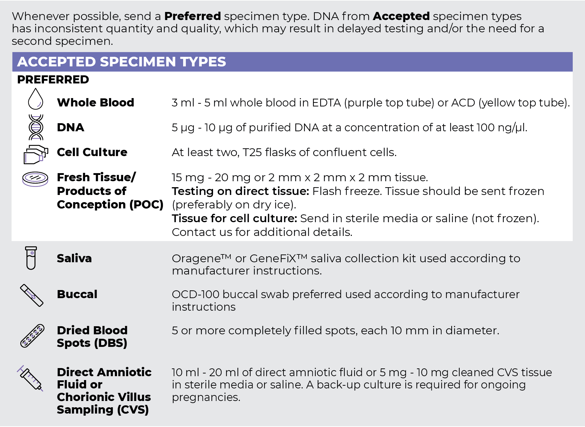
Specimen Types
Specimen Requirements and Shipping Details
PGxome (Exome) Sequencing Panel
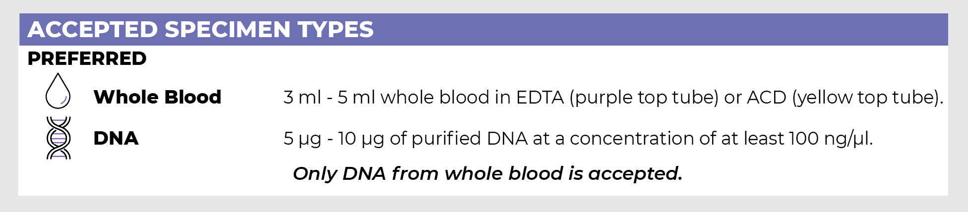
PGnome (Genome) Sequencing Panel
ORDER OPTIONS
View Ordering Instructions1) Select Test Type
2) Select Additional Test Options
No Additional Test Options are available for this test.