


Osteogenesis Imperfecta, Hypophosphatasia (HPP), and Inherited Hypophosphatemic Rickets Panel
Summary and Pricing 
Test Method
Exome Sequencing with CNV Detection| Test Code | Test Copy Genes | Panel CPT Code | Gene CPT Codes Copy CPT Code | Base Price | |
|---|---|---|---|---|---|
| 3253 | Genes x (37) | 81479 | 81404(x1), 81406(x2), 81408(x2), 81479(x69) | $990 | Order Options and Pricing |
Pricing Comments
We are happy to accommodate requests for testing single genes in this panel or a subset of these genes. The price will remain the list price. If desired, free reflex testing to remaining genes on panel is available. Alternatively, a single gene or subset of genes can also be ordered via our Custom Panel tool.
An additional 25% charge will be applied to STAT orders. STAT orders are prioritized throughout the testing process.
Click here for costs to reflex to whole PGxome (if original test is on PGxome Sequencing platform).
Click here for costs to reflex to whole PGnome (if original test is on PGnome Sequencing platform).
Turnaround Time
3 weeks on average for standard orders or 2 weeks on average for STAT orders.
Please note: Once the testing process begins, an Estimated Report Date (ERD) range will be displayed in the portal. This is the most accurate prediction of when your report will be complete and may differ from the average TAT published on our website. About 85% of our tests will be reported within or before the ERD range. We will notify you of significant delays or holds which will impact the ERD. Learn more about turnaround times here.
Targeted Testing
For ordering sequencing of targeted known variants, go to our Targeted Variants page.
Clinical Features and Genetics
Clinical Features
Osteogenesis imperfecta (OI) is a clinically and genetically heterogeneous skeletal disorder characterized by frequent bone fractures with or without minimal trauma. Clinical signs of OI can range from mild to severe. In addition to bone fractures, patients may have scoliosis, bowing of long bones, short stature, blue sclera, hearing loss, dentin defects, muscle weakness or joint laxity. Bone fractures and bowing of long bones in osteogenesis imperfecta patients may occur prenatally in severe OI cases, and hearing loss may occur in ~50% of type I OI patients by 40-years-old (https://emedicine.medscape.com/article/947588-overview). The incidence is approximately 6/100,000 (van Dijk et al. 2012. PubMed ID: 21829228). Approximately 90% of clinically diagnosed OI is caused by pathogenic variants in the COL1A1 and COL1A2 genes, while ~10% is caused by pathogenic variants in the BMP1, CREB3L1, CRTAP, FKBP10, IFITM5, P3H1(also called LEPRE1), PLOD2, PPIB, SEC24D, SERPINF1, SERPINH1, SP7, WNT1, TMEM38B, NTRK1 and other undefined genes (van Dijk and Sillence. 2014. PubMed ID: 24715559; Valadares et al. 2014. PubMed ID: 25046257; Moosa et al. 2015. PubMed ID: 26467156; Caparros-Martin et al. 2017. PubMed ID: 28116328).
Hypophosphatasia (HPP) is characterized by defective mineralization of bone and/or teeth in the presence of low activity of serum and bone alkaline phosphatase. Clinical features range from stillbirth without mineralized bone at the severe end to pathologic fractures of the lower extremities in later adulthood at the mild end. At least six clinical forms are currently recognized based on age at diagnosis and severity of features, including: (1) perinatal lethal HPP characterized by respiratory insufficiency and hypercalcemia; (2) perinatal benign HPP with prenatal skeletal manifestations that slowly resolve into the milder childhood or adult form; (3) infantile HPP with onset between birth and age six months of rickets; (4) childhood HPP that ranges from low bone mineral density for age with unexplained fractures to rickets; (5) adult HPP characterized by early loss of adult dentition and stress fractures and pseudofractures of the lower extremities in middle age; and (6) odontohypophosphatasia characterized by premature exfoliation of primary teeth and/or severe dental caries as an isolated finding or as part of the above forms of HPP (Mornet and Nunes. 2016. PubMed ID: 20301329). HPP is caused by pathogenic variants in the ALPL gene.
Hypophosphatemic rickets is a condition of abnormal phosphate homeostasis characterized by renal phosphate wasting, hypophosphatemia, and rickets/osteomalacia. Patients usually manifest bone deformity such as bowed legs after 2 years old. Patients will also develop pain in the pelvis and legs with age (Bastepe and Jüppner. 2008. PubMed ID: 18365315).
Genetics
Genes Related to Autosomal Dominant OI: Pathogenic variants in the COL1A1, COL1A2, and IFITM5 genes cause autosomal dominant OI. More than 95% of pathogenic variants in the COL1A1 and COL1A2 genes are nucleotide substitutions or small deletions or insertions, 1% -2% of COL1A1 and COL1A2 pathogenic variants are larger deletions or insertions (van Dijk and Sillence. 2014. PubMed ID: 24715559; Steiner and Basel. 2019. PubMed ID: 20301472). Almost all perinatal lethal OI are caused by de novo variants in COL1A1 and COL1A2 (Steiner and Basel. 2019. PubMed ID: 20301472).
Genes Related to Autosomal Recessive OI: Pathogenic variants in the BMP1, CREB3L1, CRTAP, FKBP10, P3H1 (also called LEPRE1), PLOD2, PPIB, SEC24D, SERPINF1, SERPINH1, SP7, TMEM38B, TENT5A, MBTPS2, NBAS, SLC2A2, SPARC, and TAPT1 genes cause autosomal recessive OI.
Recently, pathogenic variants in TENT5A (Doyard. 2018. PubMed ID: 29358272), NBAS (Balasubramanian et al. 2017. PubMed ID: 27789416), SLC2A2 (Caparros-Martin et al. 2017. PubMed ID: 28116328), SPARC (Mendoza-Londono et al. 2015. PubMed ID: 26027498), and TAPT1 (Symoens et al. 2015. PubMed ID: 26365339) have been reported in a few patients with autosomal recessive osteogenesis imperfecta.
Pathogenic variants in the XYLT2 gene have been reported in patients with autosomal recessive Spondyloocular syndrome (Taylan et al. 2016. PubMed ID: 26987875), which is characterized by spinal and long bone fractures, osteoporosis, cataract, hearing impairment, cardiac septal defects, and learning difficulties.
Genes related to both autosomal dominant and autosomal recessive OI: WNT1 pathogenic variants mainly cause autosomal recessive OI, but WNT1 has also been reported to be associated with autosomal dominant OI in rare cases.
Autosomal dominant and autosomal recessive Hypophosphatasia are caused by pathogenic variants in the ALPL gene. The majority of ALPL variants are inherited, in rare cases, de novo variants were reported (Mornet and Nunes. 2016. PubMed ID: 20301329; Taillandier et al. 2005. PubMed ID: 15671102).
Pathogenic variants in LRP5 cause autosomal recessive osteoporosis-pseudoglioma syndrome, autosomal dominant Osteopetrosis, type 1 and autosomal dominant bone mineral density variability 1.
Genes Related to X-linked OI: Pathogenic variants in PLS3 cause X-linked dominant Bone mineral density QTL18 osteoporosis.
Pathogenic variants in MBTPS2 were reported to be associated with X-linked osteogenesis imperfecta (Lindert et al. 2016. PubMed ID: 27380894).
Genes Involved in Hypophosphatemic Rickets: X-linked Hypophosphatemic Rickets is mainly caused by deleterious variants in the PHEX gene and rarely caused by deleterious variants in CLCN5 (Hauer et al. 2018. PubMed ID: 29758562). Approximately 83% of female sporadic PHEX-related X-linked Hypophosphatemic ricket cases had a de novo variant (Durmaz et al. 2013. PubMed ID: 23079138).
Autosomal dominant Hypophosphatemic Rickets is caused by pathogenic variants in FGF23 gene.
Autosomal recessive Hypophosphatemic Rickets is currently known to be caused by deleterious variants in the CYP27B1, DMP1, SLC34A3, ENPP1, CYP2R1 genes.
Pathogenic variants SLC34A3 gene also cause autosomal recessive Hypophosphatemic Rickets with hypercalcuria.
Pathogenic variants in ENPP1 also cause autosomal recessive Arterial calcification, generalized, of infancy, type 1.
Pathogenic variants in FGF23 also cause autosomal recessive Tumoral calcinosis, familial hyperphosphatemic.
Pathogenic variants in the SLC34A1 have been reported in patients with both autosomal dominant and autosomal recessive hypophosphataemic nephrolithiasis/osteoporosis, as well as hypercalcemia (Braun et al. 2016. PubMed ID: 26787776).
See individual gene summaries for more information about molecular biology of gene products and spectra of pathogenic variants.
Clinical Sensitivity - Sequencing with CNV PGxome
Pathogenic variants in COL1A1 and COL1A2 were found in 90% of individuals with Osteogenesis Imperfecta (OI) types I, II, III, or IV (Steiner and Basel. 2019. PubMed ID: 20301472). A recent study reported that COL1A1 and COL1A2 pathogenic were identified in 56% (14/35) and 44% (11/35) of the OI cases, respectively (Stephen et al. 2014. PubMed ID: 24668929).
Only one large homozygous deletion involving CREB3L1 was reported in a patient affected with autosomal recessive OI (Symoens et al. 2013. PubMed ID: 24079343).
CRTAP pathogenic variants were identified in 1 out of 10 clinically diagnosed autosomal recessive OI families (Caparrós-Martin et al. 2013. PubMed ID: 23613367).
In one publication, FKBP10 pathogenic variants were identified in 21 families affected with either autosomal recessive OI or Bruck syndrome (Schwarze et al. 2013. PubMed ID: 22949511). The pathogenic variants detection rate should be high, because all reported FKBP10 pathogenic variants are point variants or small deletions and insertions, which can be detected by sequencing.
So far, only two pathogenic IFITM5 variants have been reported (Guillén-Navarro et al. 2014. PubMed ID: 24478195). The clinical sensitivity should be high for patients with clinically diagnosed OI type V. The c.-14C>T IFITM5 pathogenic variant was found in almost all clinically diagnosed OI Type V patients tested (Lazarus et al. 2014. PubMed ID: 24674092; Human Gene Mutation Database).
P3H1 pathogenic variants were identified in 2 out of 10 clinically diagnosed autosomal recessive OI families (Caparrós-Martin et al. 2013. PubMed ID: 23613367).
SERPINF1 pathogenic variants were identified in 3 out of 10 clinically diagnosed autosomal recessive OI families (Caparrós-Martin et al. 2013. PubMed ID: 23613367).
SERPINH1 pathogenic variants were identified in 1 out of 30 individuals who tested negative for pathogenic variants in COL1A1, COL1A2, CRTAP and LEPRE1 (Christiansen et al. 2010. PubMed ID: 20188343).
So far, only one pathogenic SP7 pathogenic variant, a small deletion, has been reported (Lapunzina et al. 2010. PubMed ID: 20579626).
WNT1 pathogenic variants were reported in 5 out of 12 OI families in which no pathogenic variants in other OI-related genes were found (Keupp et al. 2013. PubMed ID: 23499309).
PLOD2 pathogenic variants were identified in 4 out of 6 clinically diagnosed, consanguineous, unrelated Egyptian families affected with Bruck syndrome type 2 (Puig-Hervás et al. 2012. PubMed ID: 22689593).
Recently, a homozygous missense variant in the NTRK1 gene has been reported in one patient with OI-related features (Caparros-Martin et al. 2017. PubMed ID: 28116328).
Sequencing of ALPL is predicted to detect pathogenic variants in 95% of cases with severe perinatal and infantile HPP. In milder forms, the detection rate is difficult to estimate. Overall, ~50% of cases with a clinical diagnosis of HPP have two ALPL pathogenic variants and 40%-45% have one pathogenic variant. The milder the disease, the higher the proportion in which only one ALPL pathogenic variant is detected (Mornet and Nunes. 2016. PubMed ID: 20301329).
To our knowledge, only a few large deletions involving ALPL have been reported (Mornet and Nunes. 2016. PubMed ID: 20301329), and large deletions account for 2.2% of reported ALPL pathogenic variants in a ALPL mutation database (http://www.sesep.uvsq.fr/03_hypo_mutations.php). Only two large deletions to date have been reported in LRP5 for osteoporosis-pseudoglioma syndrome (Human Gene Mutation Database).
Only a few DMP1 pathogenic variants have been reported (1 missense, 1 nonsense, 2 splicing, 4 small deletion and one large deletion (Farrow et al. 2009. PubMed ID: 19007919; Human Gene Mutation Database). In one study, a DMP1 pathogenic variant was identified in all three studied families with autosomal recessive hypophosphatemia (Lorenz-Depiereux et al. 2006. PubMed ID: 17033625).
Pathogenic variants in ENPP1 were found in 62 of 92 affected patients who were from 85 unrelated families with clinical diagnosed Spontaneous pathologic arterial calcifications or pseudoxanthoma elasticum (Nitschke et al. 2012. PubMed ID: 22209248). Only one large deletion including exons 24 to 25 of the ENPP1 gene was reported in a patient affected with Autosomal-Recessive Hypophosphatemic Rickets (Lorenz-Depiereux et al. 2010. PubMed ID: 20137773).
CYP27B1 pathogenic variants were reported in 22 patients from 13 Turkish families affected with Vitamin D-Dependent Rickets Type I, and the recurrent variants were c.195 + 2T>G, c.574A>G, and c.590G>A. (Tahir et al. 2016. PubMed ID: 26982175). All reported pathogenic variants were missense, small deletion/duplications, or splicing site variants. No large deletion/duplications have been reported (Human Gene Mutation Database).
A total of 15 missense pathogenic variants and one large deletion in the FGF23 gene have been reported (Human Gene Mutation Database). One large deletion involving FGF23 was reported in a family with three sibs affected with hyperphosphatemia with calcification and tumoral calcinosis, who also carried another FGF23 missense mutation (Shah et al. 2014. PubMed ID: 25378588).
In one study, PHEX pathogenic variants were identified 93 out of 118 probands (79%) (Gaucher et al. 2009. PubMed ID: 19219621). In another study, PHEX pathogenic variants were identified in 20 out of 24 unrelated probands (83%); three of these probands carried a large deletion or duplication detected by MLPA (Beck-Nielsen et al. 2012. PubMed ID: 22695891).
In one study, SLC34A3 pathogenic variants were found in one patient from 268 studied families with Nephrocalcinosis (Halbritter et al. 2015. PubMed ID: 25296721). In another study, SLC34A3 pathogenic variants were found in all 5 studied families with autosomal recessive Hypophosphatemic Rickets with Hypercalciuria (Lorenz-Depiereux et al. 2006. PubMed ID: 16358215).
Testing Strategy
This test is performed using Next-Gen sequencing with additional Sanger sequencing as necessary.
This panel typically provides 99.9% coverage of all coding exons of the genes plus 10 bases of flanking noncoding DNA in all available transcripts along with other non-coding regions in which pathogenic variants have been identified at PreventionGenetics or reported elsewhere. We define coverage as ≥20X NGS reads or Sanger sequencing. PGnome panels typically provide slightly increased coverage over the PGxome equivalent. PGnome sequencing panels have the added benefit of additional analysis and reporting of deep intronic regions (where applicable).
Dependent on the sequencing backbone selected for this testing, discounted reflex testing to any other similar backbone-based test is available (i.e., PGxome panel to whole PGxome; PGnome panel to whole PGnome).
Indications for Test
Candidates for this test include patients with a clinical presentation of Osteogenesis imperfecta, or hypophosphatasia, or Inherited Hypophosphatemic Rickets.
Candidates for this test include patients with a clinical presentation of Osteogenesis imperfecta, or hypophosphatasia, or Inherited Hypophosphatemic Rickets.
Genes
| Inheritance | Abbreviation |
|---|---|
| Autosomal Dominant | AD |
| Autosomal Recessive | AR |
| X-Linked | XL |
| Mitochondrial | MT |
Diseases
Related Test
| Name |
|---|
| PGxome® |
Citations
- Balasubramanian et al. 2017. PubMed ID: 27789416
- Bastepe and Jüppner. 2008. PubMed ID: 18365315
- Beck-Nielsen et al. 2012. PubMed ID: 22695891
- Braun et al. 2016. PubMed ID: 26787776
- Caparrós-Martin et al. 2013. PubMed ID: 23613367
- Caparros-Martin et al. 2017. PubMed ID: 28116328
- Christiansen et al. 2010. PubMed ID: 20188343
- Doyard. 2018. PubMed ID: 29358272
- Durmaz et al. 2013. PubMed ID: 23079138
- Farrow et al. 2009. PubMed ID: 19007919
- Gaucher et al. 2009. PubMed ID: 19219621
- Guillén-Navarro et al. 2014. PubMed ID: 24478195
- Halbritter et al. 2015. PubMed ID: 25296721
- Hauer et al. 2018. PubMed ID: 29758562
- Human Gene Mutation Database (Bio-base).
- Keupp et al. 2013. PubMed ID: 23499309
- Lapunzina et al. 2010. PubMed ID: 20579626
- Lazarus et al. 2014. PubMed ID: 24674092
- Lindert et al. 2016. PubMed ID: 27380894
- Lorenz-Depiereux et al. 2006. PubMed ID: 17033625
- Lorenz-Depiereux et al. 2006. PubMed ID: 16358215
- Lorenz-Depiereux et al. 2010. PubMed ID: 20137773
- Mendoza-Londono et al. 2015. PubMed ID: 26027498
- Moosa et al. 2015. PubMed ID: 26467156
- Mornet and Nunes. 2016. PubMed ID: 20301329
- Nitschke et al. 2012. PubMed ID: 22209248
- Puig-Hervás et al. 2012. PubMed ID: 22689593
- Schwarze et al. 2013. PubMed ID: 22949511
- Shah et al. 2014. PubMed ID: 25378588
- Steiner and Basel. 2019. PubMed ID: 20301472
- Stephen et al. 2014. PubMed ID: 24668929
- Symoens et al. 2013. PubMed ID: 24079343
- Symoens et al. 2015. PubMed ID: 26365339
- Tahir et al. 2016. PubMed ID: 26982175
- Taillandier et al. 2005. PubMed ID: 15671102
- Taylan et al. 2016. PubMed ID: 26987875
- Valadares et al. 2014. PubMed ID: 25046257
- van Dijk and Sillence. 2014. PubMed ID: 24715559
- van Dijk et al. 2012. PubMed ID: 21829228
Ordering/Specimens
Ordering Options
We offer several options when ordering sequencing tests. For more information on these options, see our Ordering Instructions page. To view available options, click on the Order Options button within the test description.
myPrevent - Online Ordering
- The test can be added to your online orders in the Summary and Pricing section.
- Once the test has been added log in to myPrevent to fill out an online requisition form.
- PGnome sequencing panels can be ordered via the myPrevent portal only at this time.
Requisition Form
- A completed requisition form must accompany all specimens.
- Billing information along with specimen and shipping instructions are within the requisition form.
- All testing must be ordered by a qualified healthcare provider.
For Requisition Forms, visit our Forms page
If ordering a Duo or Trio test, the proband and all comparator samples are required to initiate testing. If we do not receive all required samples for the test ordered within 21 days, we will convert the order to the most effective testing strategy with the samples available. Prior authorization and/or billing in place may be impacted by a change in test code.
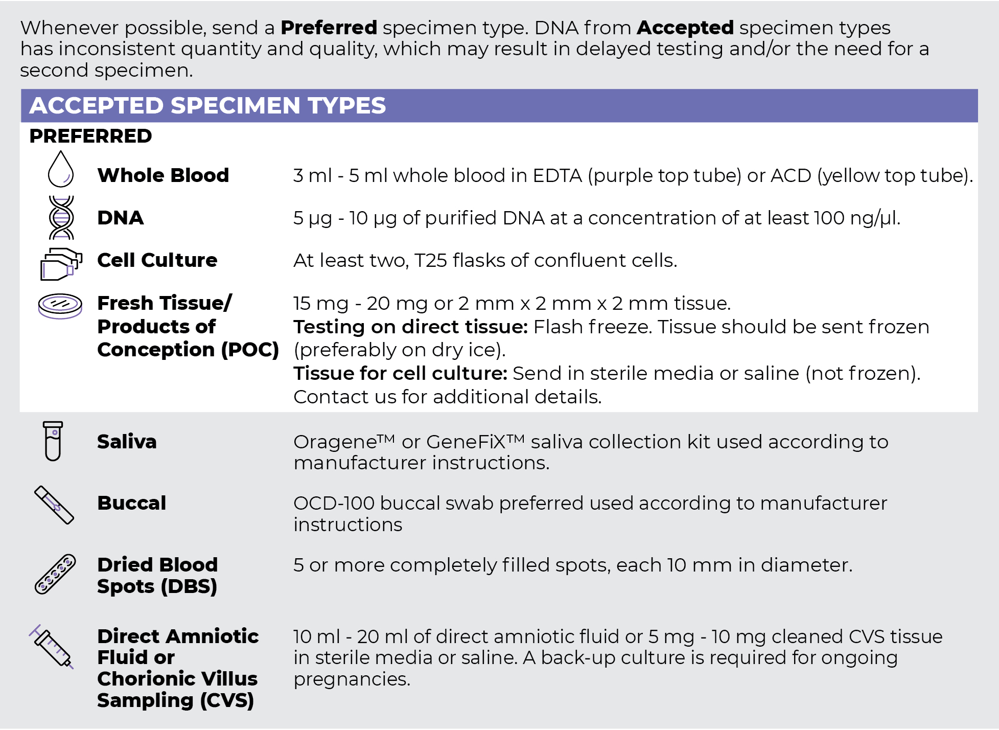
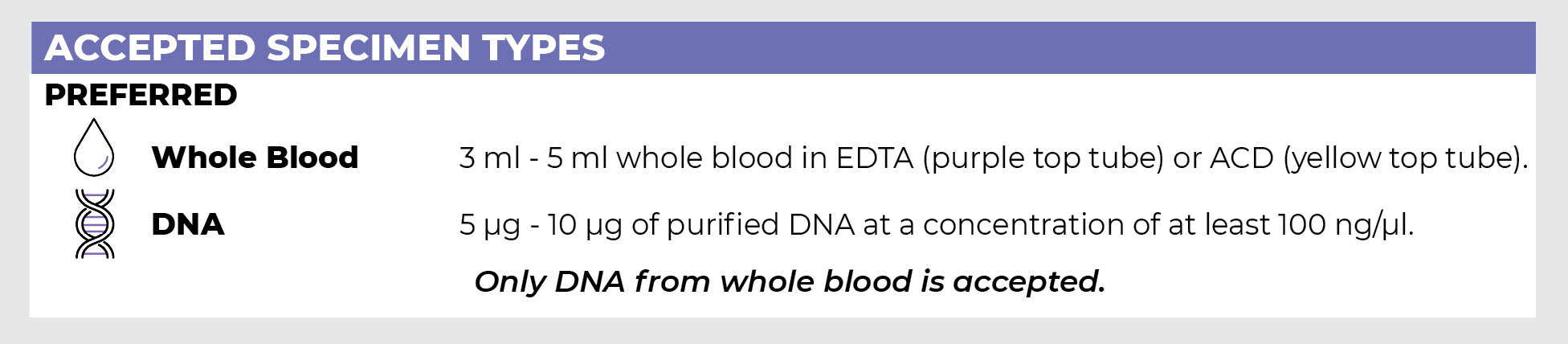
Specimen Types
Specimen Requirements and Shipping Details
PGxome (Exome) Sequencing Panel
PGnome (Genome) Sequencing Panel
ORDER OPTIONS
View Ordering Instructions1) Select Test Type
2) Select Additional Test Options
No Additional Test Options are available for this test.