


Infantile Myofibromatosis and Idiopathic Basal Ganglia Calcification via the PDGFRB Gene
Summary and Pricing 
Test Method
Exome Sequencing with CNV Detection| Test Code | Test Copy Genes | Test CPT Code | Gene CPT Codes Copy CPT Code | Base Price | |
|---|---|---|---|---|---|
| 11563 | PDGFRB | 81479 | 81479,81479 | $990 | Order Options and Pricing |
Pricing Comments
Our favored testing approach is exome based NextGen sequencing with CNV analysis. This will allow cost effective reflexing to PGxome or other exome based tests. However, if full gene Sanger sequencing is desired for STAT turnaround time, insurance, or other reasons, please see link below for Test Code, pricing, and turnaround time information.
An additional 25% charge will be applied to STAT orders. STAT orders are prioritized throughout the testing process.
Click here for costs to reflex to whole PGxome (if original test is on PGxome Sequencing platform).
Click here for costs to reflex to whole PGnome (if original test is on PGnome Sequencing platform).
The Sanger Sequencing method for this test is NY State approved.
For Sanger Sequencing click here.Turnaround Time
3 weeks on average for standard orders or 2 weeks on average for STAT orders.
Please note: Once the testing process begins, an Estimated Report Date (ERD) range will be displayed in the portal. This is the most accurate prediction of when your report will be complete and may differ from the average TAT published on our website. About 85% of our tests will be reported within or before the ERD range. We will notify you of significant delays or holds which will impact the ERD. Learn more about turnaround times here.
Targeted Testing
For ordering sequencing of targeted known variants, go to our Targeted Variants page.
Clinical Features and Genetics
Clinical Features
Infantile myofibromatosis (IM), previously known as congenital generalized fibromatosis, is characterized by the development of tumors in various tissues and organs. IM affects mostly infants and young children. Although tumors are usually detected at birth or during the first two years of life, uterine and adult onsets have been also reported (Chung and Enzinger 1981).
Two main types of IM, solitary and multicentric, are distinguished. Each type is further divided in two groups based on the presence or absence of visceral involvement (Wiswell et al. 1988).
The solitary type is characterized by the development of a single nodule mainly in the bones, striated muscle, skin, or subcutaneous tissues. In rare cases, solitary tumors have been reported in the viscera.
The multicentric type is characterized by the development of multiple nodules in various organs. Visceral involvement is common in this type. The lungs, heart, gastrointestinal tract, pancreas, liver, and bones are most commonly affected. Involvement of the central nervous system appears to be rare (Wada et al. 1998). IM with visceral involvement has a poor prognosis, although spontaneous regression has been reported in some cases (Teng et al. 1963).
About half the patients with IM have the solitary type and the other half have the multicentric type. The solitary type affects older individuals in about 20% of patients. Both solitary and multicentric IM have a good prognosis in the absence of visceral involvement when the tumors regress spontaneously during the first years of life. However, new tumors may develop later.
IM is clinically heterogeneous. Symptoms are highly variable and depend on the number and location of tumors. The disease may be limited to the skin in some patients, while several visceral organs are involved in other patients. Affected related members may present either with the solitary type with no visceral involvement or the multicentric visceral type (Cheung et al. 2013). The tumors consist of soft tissues abnormalities, and are usually benign. However, complications may occur when the tumors affect the normal function of vital organs such as the brain or viscera.
Although rare, IM has been reported in various ethnic and geographical groups (Cheung et al. 2013).
Familial Idiopathic Basal Ganglia Calcification (IBGC), also known as Fahr’s syndrome, is a neurological disorder characterized by abnormal deposits of calcium in areas of the brain that control movement. The radiological characteristics of IBGC consist of bilateral and symmetrical calcification of the basal ganglia. Additional areas of the brain may also be affected. IBGC is clinically heterogeneous. Symptoms usually begin during the fourth and fifth decade of life. Childhood and adolescent onset have been also reported. Movement disorders in the form of dystonia, tremor and chorea are the initial and most common clinical manifestations of IBGC. As the disease progress neurological and neuropsychiatric abnormalities are detected and include seizures, spasticity, headache, dysarthria, psychosis, mood disturbances, cognitive decline, and dementia (Manyam 2005; Nicolas et al. 2013b).
Abnormal deposit of calcium in the brain is a common finding usually associated with aging. However, familial IBGC is rare, with less than 1/1,000,000 people affected worldwide (Ellie et al. 1989).
Genetics
In most of the IM-affected families reported, the disease is inherited in an autosomal dominant manner. In rare families, a recessive mode of transmission is speculated. It has been argued that family history may be difficult to obtain due to the spontaneous regression of tumors (Narchi 2001).
Autosomal Dominant IM is genetically heterogeneous. Two genes, PDGFRB and NOTCH3 have been recently implicated in the disease. Two germline missense mutations in the PDGFRB gene, c.1681C>T (p.Arg561Cys) and c.1978C>A (p.Pro660Thr) have been reported. The c.1681C>T variant was reported in families from different ethnic and geographical populations (Cheng et al. 2013; Martignetti et al. 2013).
Familial IBGC is a genetically heterogeneous autosomal dominant disorder. Simplex cases with no apparent family history have been also reported. To date, three genes have been associated with the disease: SLC20A2, PDGFB, and PDGFRB. Three pathogenic missense variants in PDGFRB have been recently reported both in familial and apparently simplex cases (Nicolas 2013a; 2013b).
PDGFRB encodes the platelet-derived growth factor receptor beta, a tyrosine kinase receptor that has been involved in several cellular processes including proliferation, differentiation, survival, and migration. It has also been suggested that the PDGFRB/ PDGFB pathway is involved in the calcification of vascular smooth muscle cells (Diliberto et al. 1991).
Clinical Sensitivity - Sequencing with CNV PGxome
Pathogenic variants in the PDGFRB gene are the major cause of familial IM; they were found in 12 out of 13 IM-affected families (Cheng et al. 2013; Martignetti et al. 2013). This test will detect PDGFRB pathogenic variants in ~ 4% of patients with IBGC (Nicolas et al. 2013b).
Testing Strategy
This test provides full coverage of all coding exons of the PDGFRB gene plus 10 bases of flanking noncoding DNA in all available transcripts along with other non-coding regions in which pathogenic variants have been identified at PreventionGenetics or reported elsewhere. We define full coverage as >20X NGS reads or Sanger sequencing. PGnome panels typically provide slightly increased coverage over the PGxome equivalent. PGnome sequencing panels have the added benefit of additional analysis and reporting of deep intronic regions (where applicable).
Dependent on the sequencing backbone selected for this testing, discounted reflex testing to any other similar backbone-based test is available (i.e., PGxome panel to whole PGxome; PGnome panel to whole PGnome).
Indications for Test
Patients with clinical and radiological findings suggestive of infantile myofibromatosis or Idiopathic Basal Ganglia Calcification are candidates.
Patients with clinical and radiological findings suggestive of infantile myofibromatosis or Idiopathic Basal Ganglia Calcification are candidates.
Gene
| Official Gene Symbol | OMIM ID |
|---|---|
| PDGFRB | 173410 |
| Inheritance | Abbreviation |
|---|---|
| Autosomal Dominant | AD |
| Autosomal Recessive | AR |
| X-Linked | XL |
| Mitochondrial | MT |
Diseases
| Name | Inheritance | OMIM ID |
|---|---|---|
| Basal Ganglia Calcification, Idiopathic, 4 | AD | 615007 |
| Myofibromatosis, Infantile, 1 | AD | 228550 |
Citations
- Cheung YH, Gayden T, Campeau PM, LeDuc CA, Russo D, Nguyen V-H, Guo J, Qi M, Guan Y, Albrecht S, Moroz B, Eldin KW, et al. 2013. A recurrent PDGFRB mutation causes familial infantile myofibromatosis. Am. J. Hum. Genet. 92: 996–1000. PubMed ID: 23731537
- Chung EB, Enzinger FM. 1981. Infantile myofibromatosis. Cancer 48: 1807–1818. PubMed ID: 7284977
- Diliberto PA, Gordon G, Herman B. 1991. Regional and mechanistic differences in platelet-derived growth factor-isoform-induced alterations in cytosolic free calcium in porcine vascular smooth muscle cells. J. Biol. Chem. 266: 12612–12617. PubMed ID: 1905727
- Ellie E, Julien J, Ferrer X. 1989. Familial idiopathic striopallidodentate calcifications. Neurology 39: 381–385. PubMed ID: 2927646
- Manyam BV. 2005. What is and what is not “Fahr”s disease’. Parkinsonism Relat. Disord. 11: 73–80. PubMed ID: 15734663
- Martignetti JA, Tian L, Li D, Ramirez MCM, Camacho-Vanegas O, Camacho SC, Guo Y, Zand DJ, Bernstein AM, Masur SK, Kim CE, Otieno FG, et al. 2013. Mutations in PDGFRB Cause Autosomal-Dominant Infantile Myofibromatosis. The American Journal of Human Genetics 92: 1001–1007. PubMed ID: 23731542
- Narchi H. 2001. Four half-siblings with infantile myofibromatosis: a case for autosomal-recessive inheritance. Clin. Genet. 59: 134–135. PubMed ID: 11260217
- Nicolas G, Pottier C, Charbonnier C, Guyant-Maréchal L, Ber I Le, Pariente J, Labauge P, Ayrignac X, Defebvre L, Maltête D, Martinaud O, Lefaucheur R, et al. 2013b. Phenotypic spectrum of probable and genetically-confirmed idiopathic basal ganglia calcification. Brain 136: 3395–3407. PubMed ID: 24065723
- Nicolas G, Pottier C, Maltête D, Coutant S, Rovelet-Lecrux A, Legallic S, Rousseau S, Vaschalde Y, Guyant-Maréchal L, Augustin J, Martinaud O, Defebvre L, et al. 2013a. Mutation of the PDGFRB gene as a cause of idiopathic basal ganglia calcification. Neurology 80: 181–187. PubMed ID: 23255827
- Teng P, Warden MJ, Cohn WL. 1963. Congenital generalized fibromatosis (renal and skeletal) with complete spontaneous regression. J. Pediatr. 62: 748–753. PubMed ID: 13980570
- Wada H, Akiyama H, Seki H, Ichihara T, Ueno K, Miyawaki T, Koizumi S. 1998. Spinal canal involvement in infantile myofibromatosis: case report and review of the literature. J. Pediatr. Hematol. Oncol. 20: 353–356. PubMed ID: 9703012
- Wiswell TE, Davis J, Cunningham BE, Solenberger R, Thomas PJ. 1988. Infantile myofibromatosis: the most common fibrous tumor of infancy. J. Pediatr. Surg. 23: 315–318. PubMed ID: 3385581
Ordering/Specimens
Ordering Options
We offer several options when ordering sequencing tests. For more information on these options, see our Ordering Instructions page. To view available options, click on the Order Options button within the test description.
myPrevent - Online Ordering
- The test can be added to your online orders in the Summary and Pricing section.
- Once the test has been added log in to myPrevent to fill out an online requisition form.
- PGnome sequencing panels can be ordered via the myPrevent portal only at this time.
Requisition Form
- A completed requisition form must accompany all specimens.
- Billing information along with specimen and shipping instructions are within the requisition form.
- All testing must be ordered by a qualified healthcare provider.
For Requisition Forms, visit our Forms page
If ordering a Duo or Trio test, the proband and all comparator samples are required to initiate testing. If we do not receive all required samples for the test ordered within 21 days, we will convert the order to the most effective testing strategy with the samples available. Prior authorization and/or billing in place may be impacted by a change in test code.
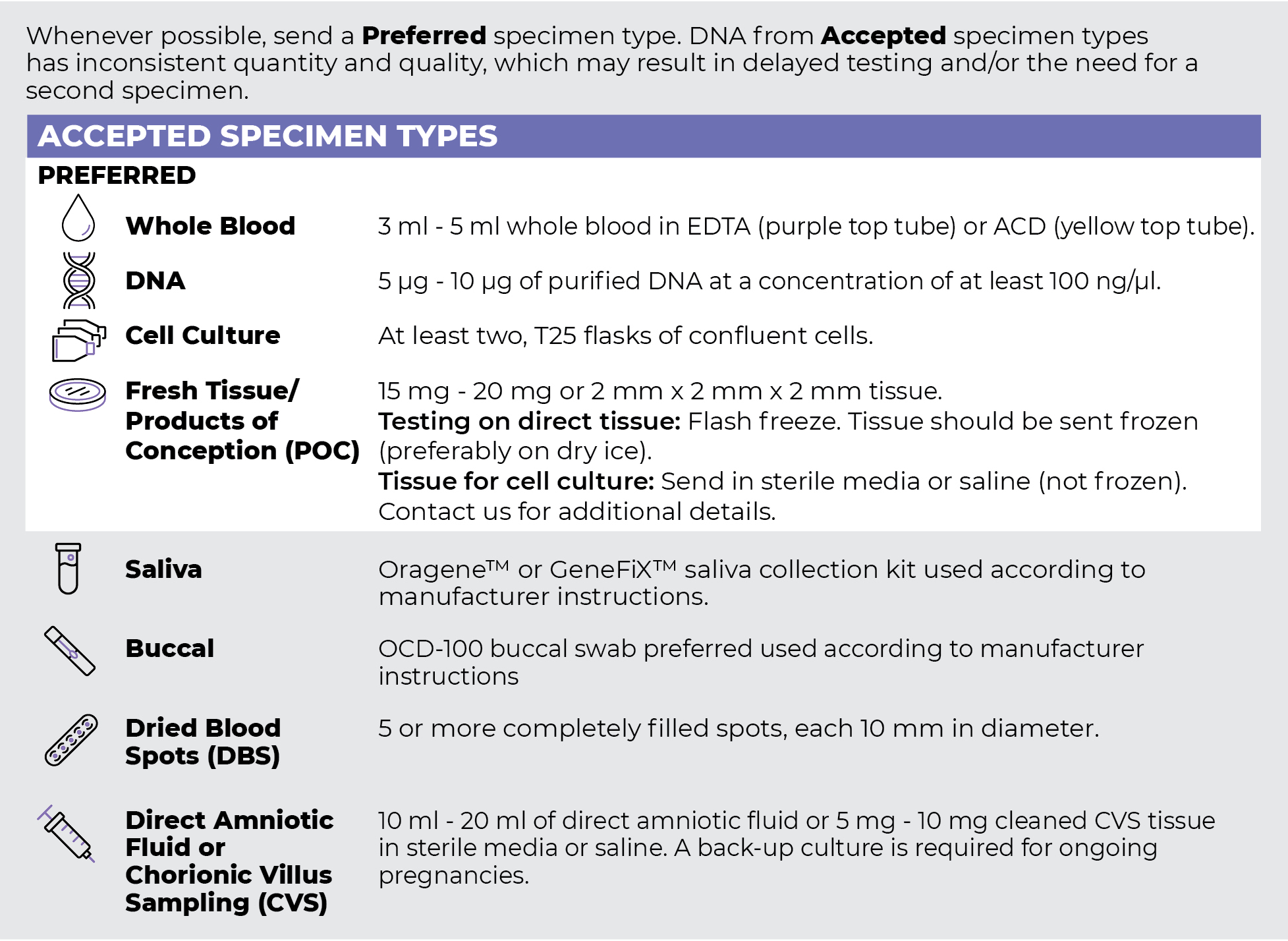
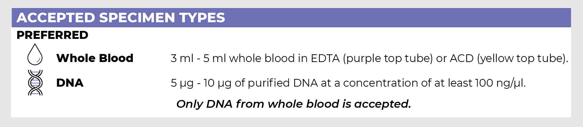
Specimen Types
Specimen Requirements and Shipping Details
PGxome (Exome) Sequencing Panel
PGnome (Genome) Sequencing Panel
ORDER OPTIONS
View Ordering Instructions1) Select Test Type
2) Select Additional Test Options
No Additional Test Options are available for this test.