


Hereditary Spherocytosis via the SLC4A1 Gene
Summary and Pricing 
Test Method
Exome Sequencing with CNV Detection| Test Code | Test Copy Genes | Test CPT Code | Gene CPT Codes Copy CPT Code | Base Price | |
|---|---|---|---|---|---|
| 11683 | SLC4A1 | 81479 | 81479,81479 | $990 | Order Options and Pricing |
Pricing Comments
Our favored testing approach is exome based NextGen sequencing with CNV analysis. This will allow cost effective reflexing to PGxome or other exome based tests. However, if full gene Sanger sequencing is desired for STAT turnaround time, insurance, or other reasons, please see link below for Test Code, pricing, and turnaround time information. If the Sanger option is selected, CNV detection may be ordered through Test #600.
An additional 25% charge will be applied to STAT orders. STAT orders are prioritized throughout the testing process.
Click here for costs to reflex to whole PGxome (if original test is on PGxome Sequencing platform).
Click here for costs to reflex to whole PGnome (if original test is on PGnome Sequencing platform).
The Sanger Sequencing method for this test is NY State approved.
For Sanger Sequencing click here.Turnaround Time
3 weeks on average for standard orders or 2 weeks on average for STAT orders.
Please note: Once the testing process begins, an Estimated Report Date (ERD) range will be displayed in the portal. This is the most accurate prediction of when your report will be complete and may differ from the average TAT published on our website. About 85% of our tests will be reported within or before the ERD range. We will notify you of significant delays or holds which will impact the ERD. Learn more about turnaround times here.
Targeted Testing
For ordering sequencing of targeted known variants, go to our Targeted Variants page.
Clinical Features and Genetics
Clinical Features
Hereditary Spherocytosis (HS), also known as Minkowski-Chauffard disease, affects one in 2,000 individuals. HS is a condition where red blood cells lose their typical biconcave disc shape and appear spherical. The spherical appearance impairs membrane flexibility making it hard for red blood cells to transverse narrow capillaries, especially in the spleen. This impairment causes anemia due to chronic extravascular hemolysis, jaundice, formation of bilirubin gallstones, reticulocytosis and splenomegaly characteristic of HS disease (Aster et al. 2013; An and Mohandas 2008). Disease severity can range with 20-30% having mild form, 60-70% having moderate form, and 10-20% having severe form of HS. People with mild forms may be asymptomatic whereas severe forms of the disease present in newborns with life threatening anemia and require blood transfusions. There are five types of HS defined by the gene mutation causative for disease: Type I-ANK1, type 2-SPTB, type 3-SPTA1, type 4-SLC4A1, and type 5-EPB42 (Bolton-Maggs et al. 2004; Delaunay 2007). Type 4 HS makes up 15-20% of cases with patients presenting with mainly with mild to moderate anemia. A few cases have been associated with severe anemia in type 4 HS (Jarolim et al 1996; Dhermy et al. 1997). Mushroomed-shaped or “pincered” red cells may also appear in addition to spherocytes in peripheral blood smears in type 4 HS (An and Mohandas 2008).
Mutations in the SLCA41 gene have been shown to cause both ovalocytosis and distal renal tubular acidosis. Ovalocytosis is predominant in southeastern-asian populations and is a form of hereditary elliptocytosis that is often asymptomatic. A few affected individuals have been reported to have varying degrees of hemolysis (Reardon et al. 1993; Coetzer et al. 1996). Distal renal tubular acidosis occurs when kidneys fail to remove acids from blood in the urine. Common symptoms include fatigue, kidney stones, nephrocalcinosis, impaired growth, rickets, and muscle weakness (Fry and Karet 2007).
Genetics
HS in inherited in an autosomal dominant manner in 75% of cases through mutations in the ANK1, SPTB, and SLC4A1 genes. Autosomal recessive forms are inherited through mutations in the ANK1, SPTA1, and EPB42 genes (Bolton-Maggs et al. 2004). Most mutations reported to date are private with de novo dominant mutations being six times more common than recessive mutations (Miraglia del Giudice et al. 2001). Mutations in the ANK1, SPTB, SLC4A1, SPTA1, and EPB42 genes account for 60%, 10%, 15%, 10%, and 5% cases of HS respectively (An and Mohandas 2008).
HS type 4 is inherited through an autosomal dominant manner through mutations in the SLC4A1 gene. Missense changes represent over half of the causative mutations in the SLC4A1 gene and can disrupt binding to membrane skeleton proteins, or prevent incorporation of Band 3 protein into the plasma membrane. Nonsense, splice site mutations and small indels leading to loss of Band 3 protein have also been described (Tse and Lux 1999). To date, no gross deletions have been reported in the SLC4A1 gene. Southeast Asian ovalocytosis is due to a 27 bp inframe deletion defined as c.1198_1224 (p.Ala400_Ala408) residing at the cytoplasmic/transmembrane domain of Band 3 (Jarolim et al. 1999). Distal renal tubular acidosis is similarly caused by missense mutation in the SLC4A1 gene non-overlapping with known HS missense mutations (Bruce et al. 1997; Tanphaichitr et al. 1998). The SLC4A1 gene encodes the Band 3 protein, an anion exchanger mediating exchange of chloride for bicarbonate across the plasma membrane. Band 3 protein also links the plasma membrane to the spectrin based cytoskeleton through cytoplasmic interaction with ankryin. This interaction network is essential in regulation cohesion between the plasma membrane and underlying cytoskeletion providing red blood cell flexibility when navigating narrow vasculature (An and Mohandas 2008).
Clinical Sensitivity - Sequencing with CNV PGxome
Mutations in the SLC4A1 gene are responsible for 15-20% of HS cases. SLC4A1 is the only known gene involved with ovalocytosis and autosomal dominant distal renal tubular acidosis (An and Mohandas 2008; Fry and Karet 2007). Analytical sensitivity is high as all mutations reported to date are detectable by sequencing.
Testing Strategy
This test provides full coverage of all coding exons of the SLC4A1 gene plus 10 bases of flanking noncoding DNA in all available transcripts along with other non-coding regions in which pathogenic variants have been identified at PreventionGenetics or reported elsewhere. We define full coverage as >20X NGS reads or Sanger sequencing. PGnome panels typically provide slightly increased coverage over the PGxome equivalent. PGnome sequencing panels have the added benefit of additional analysis and reporting of deep intronic regions (where applicable).
Dependent on the sequencing backbone selected for this testing, discounted reflex testing to any other similar backbone-based test is available (i.e., PGxome panel to whole PGxome; PGnome panel to whole PGnome).
Indications for Test
Candidates for this test are patients showing features consistent with HS (Spherocytes in peripheral blood smears, anemia and reticulocytosis) and a family history for the disorder. Other typical pathological features include increased MCHC, increased RDW, and heightened sensitivity via osmotic fragility test. HS may be differentiated between autoimmune and alloimmune hemolytic anemia via a negative Coombs test (Aster et al. 2013).
Candidates for this test are patients showing features consistent with HS (Spherocytes in peripheral blood smears, anemia and reticulocytosis) and a family history for the disorder. Other typical pathological features include increased MCHC, increased RDW, and heightened sensitivity via osmotic fragility test. HS may be differentiated between autoimmune and alloimmune hemolytic anemia via a negative Coombs test (Aster et al. 2013).
Gene
| Official Gene Symbol | OMIM ID |
|---|---|
| SLC4A1 | 109270 |
| Inheritance | Abbreviation |
|---|---|
| Autosomal Dominant | AD |
| Autosomal Recessive | AR |
| X-Linked | XL |
| Mitochondrial | MT |
Diseases
| Name | Inheritance | OMIM ID |
|---|---|---|
| Ovalocytosis | 109270 | |
| Renal Tubular Acidosis, Distal, Autosomal Dominant | AD | 179800 |
| Spherocytosis, Type 4 | AD | 612653 |
Citations
- An X, Mohandas N. 2008. Disorders of red cell membrane. Br. J. Haematol. 141: 367–375. PubMed ID: 18341630
- Aster, JC, Pozdnyakova, O, Kutok, JL. Hematopathology. Philadelphia: Elsevier Saunders, 2013.
- Bolton-Maggs PHB, Langer JC, Iolascon A, Tittensor P, King M-J, General Haematology Task Force of the British Committee for Standards in Haematology. 2012. Guidelines for the diagnosis and management of hereditary spherocytosis--2011 update. Br. J. Haematol. 156: 37–49. PubMed ID: 22055020
- Bruce LJ, Cope DL, Jones GK, Schofield AE, Burley M, Povey S, Unwin RJ, Wrong O, Tanner MJ. 1997. Familial distal renal tubular acidosis is associated with mutations in the red cell anion exchanger (Band 3, AE1) gene. J. Clin. Invest. 100: 1693–1707. PubMed ID: 9312167
- Coetzer TL, Beeton L, Zyl D van, Field SP, Agherdien A, Smart E, Daniels GL. 1996. Southeast Asian ovalocytosis in a South African kindred with hemolytic anemia. Blood 87: 1656–1657. PubMed ID: 8608262
- Delaunay J. 2007. The molecular basis of hereditary red cell membrane disorders. Blood Rev. 21: 1–20. PubMed ID: 16730867
- Dhermy D, Galand C, Bournier O, Boulanger L, Cynober T, Schismanoff PO, Bursaux E, Tchernia G, Boivin P, Garbarz M. 1997. Heterogenous band 3 deficiency in hereditary spherocytosis related to different band 3 gene defects. Br. J. Haematol. 98: 32–40. PubMed ID: 9233560
- Fry AC, Karet FE. 2007. Inherited renal acidoses. Physiology (Bethesda) 22: 202–211. PubMed ID: 17557941
- Jarolim P, Murray JL, Rubin HL, Taylor WM, Prchal JT, Ballas SK, Snyder LM, Chrobak L, Melrose WD, Brabec V, Palek J. 1996. Characterization of 13 novel band 3 gene defects in hereditary spherocytosis with band 3 deficiency. Blood 88: 4366–4374. PubMed ID: 8943874
- Miraglia del Giudice E, Nobili B, Francese M, D’Urso L, Iolascon A, Eber S, Perrotta S. 2001. Clinical and molecular evaluation of non-dominant hereditary spherocytosis. Br. J. Haematol. 112: 42–47. PubMed ID: 11167781
- Reardon DM, Seymour CA, Cox TM, Pinder JC, Schofield AE, Tanner MJ. 1993. Hereditary ovalocytosis with compensated haemolysis. Br. J. Haematol. 85: 197–199. PubMed ID: 8251392
- Tanphaichitr VS, Sumboonnanonda A, Ideguchi H, Shayakul C, Brugnara C, Takao M, Veerakul G, Alper SL. 1998. Novel AE1 mutations in recessive distal renal tubular acidosis. Loss-of-function is rescued by glycophorin A. J. Clin. Invest. 102: 2173–2179. PubMed ID: 9854053
- Tse WT, Lux SE. 1999. Red blood cell membrane disorders. Br. J. Haematol. 104: 2–13. PubMed ID: 10027705
Ordering/Specimens
Ordering Options
We offer several options when ordering sequencing tests. For more information on these options, see our Ordering Instructions page. To view available options, click on the Order Options button within the test description.
myPrevent - Online Ordering
- The test can be added to your online orders in the Summary and Pricing section.
- Once the test has been added log in to myPrevent to fill out an online requisition form.
- PGnome sequencing panels can be ordered via the myPrevent portal only at this time.
Requisition Form
- A completed requisition form must accompany all specimens.
- Billing information along with specimen and shipping instructions are within the requisition form.
- All testing must be ordered by a qualified healthcare provider.
For Requisition Forms, visit our Forms page
If ordering a Duo or Trio test, the proband and all comparator samples are required to initiate testing. If we do not receive all required samples for the test ordered within 21 days, we will convert the order to the most effective testing strategy with the samples available. Prior authorization and/or billing in place may be impacted by a change in test code.
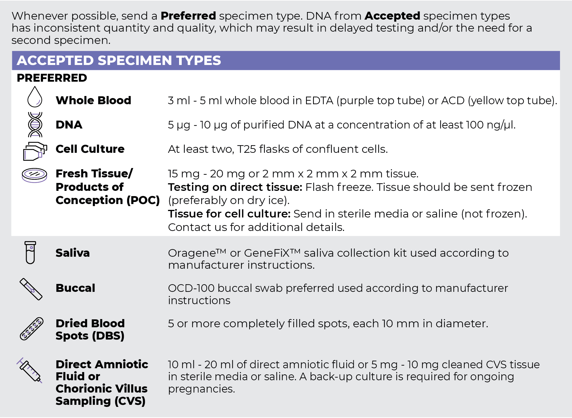
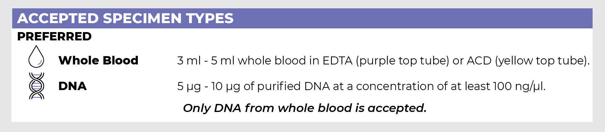
Specimen Types
Specimen Requirements and Shipping Details
PGxome (Exome) Sequencing Panel
PGnome (Genome) Sequencing Panel
ORDER OPTIONS
View Ordering Instructions1) Select Test Type
2) Select Additional Test Options
No Additional Test Options are available for this test.