


Hereditary Polyposis Panel
Summary and Pricing 
Test Method
Sequencing and CNV Detection via NextGen Sequencing using PG-Select Capture Probes| Test Code | Test Copy Genes | Panel CPT Code | Gene CPT Codes Copy CPT Code | Base Price | |
|---|---|---|---|---|---|
| 5465 | Genes x (7) | 81479 | 81201(x1), 81203(x1), 81406(x1), 81479(x11) | $990 | Order Options and Pricing |
Pricing Comments
Testing run on PG-select capture probes includes CNV analysis for the gene(s) on the panel but does not permit the optional add on of exome-wide CNV analysis. Any of the NGS platforms allow reflex to other clinically relevant genes, up to whole exome or whole genome sequencing depending upon the base platform selected for the initial test.
An additional 25% charge will be applied to STAT orders. STAT orders are prioritized throughout the testing process.
This test is also offered via a custom panel (click here) on our exome or genome backbone which permits the optional add on of exome-wide CNV or genome-wide SV analysis.
Turnaround Time
3 weeks on average for standard orders or 2 weeks on average for STAT orders.
Please note: Once the testing process begins, an Estimated Report Date (ERD) range will be displayed in the portal. This is the most accurate prediction of when your report will be complete and may differ from the average TAT published on our website. About 85% of our tests will be reported within or before the ERD range. We will notify you of significant delays or holds which will impact the ERD. Learn more about turnaround times here.
Targeted Testing
For ordering sequencing of targeted known variants, go to our Targeted Variants page.
Clinical Features and Genetics
Clinical Features
Colorectal cancer (CRC) is defined by development of tumors in the colon and rectum. CRC is generally classified by the presence or absence of polyposis (numerous internal polyps) and has been categorized into specific inherited diseases based on the degree of polyposis and other physiologically-defining features. CRC includes Lynch syndrome (also known as hereditary non-polyposis CRC), which is defined by age of onset of cancer before age 50 and delineated by specific criteria (Amsterdam Criteria) for diagnosis (Vasen et al. 1999. PubMed ID: 10348829). Peutz-Jeghers syndrome, Cowden syndrome, and juvenile polyposis are each associated with increased risk of hereditary cancers (such as CRC) but also present with additional physiological anomalies (Eng et al. 2014. PubMed ID: 20301661; Eng et al. 2001. PubMed ID: 11160785; Abdalla et al. 2003. PubMed ID: 12843319; Hearle et al. 2006. PubMed ID: 16707622).
Familial adenomatous polyposis (FAP) and MUTYH–associated polyposis (MAP) are specifically associated with an abundance of internal polyps that have the potential to progress to CRC. Thirty percent of CRCs are considered familial, while 5% are caused by a Mendelian disorder (Esteban-Jurado 2014. PubMed ID: 24587672). Early surveillance and treatment have been shown to decrease morbidity and mortality, and identification of pathogenic variants is important for determining the degree of cancer surveillance (colonoscopy) necessary for affected individuals and family members (Kohlmann and Gruber. 2018. PubMed ID: 20301390).
Familial adenomatous polyposis (FAP) is characterized by polyposis of the colon (>100 polyps) that begins to form around the age of 16. FAP accounts for 1% of all CRCs (Hegde et al. 2014. PubMed ID: 24310308). If untreated, nearly all FAP patients will develop CRC by age 40 (Fearnhead et al. 2001. PubMed ID: 11257105). Individuals with FAP can also present with desmoid tumors, congenital hypertrophy of retinal pigment epithelium (CHRPE; 60% of individuals), osteomas, supernumerary teeth, odontomas, epidermoid cysts, and small bowel adenomas (Hegde et al. 2014. PubMed ID: 24310308). CHRPE does not affect sight or have malignant potential but can be detected by ophthalmoscopy and is highly diagnostic of FAP before the appearance of polyps (Díaz-Llopis and Menezo. 1988. PubMed ID: 2830869).
MAP is a specific form of FAP in which individuals present by age 55 with colorectal adenomas (10-1000; Poulsen and Bisgaard. 2008. PubMed ID: 19506731). Polyp burden in these cases is variable, as biallelic variants in MUTYH have been found in 30% of individuals with 15-100 polyps, and 7% of individuals with >100 polyps. Due to the function of MUTYH in the base excision repair system, colorectal adenomas and carcinomas from these patients include numerous G:C to T:A transversions in various genes such as APC and KRAS (K-ras tumor suppressor; Lipton et al. 2003. PubMed ID: 14633673; Jones et al. 2004. PubMed ID: 15083190; Hegde et al. 2014. PubMed ID: 24310308).
Genetics
The APC gene is best characterized for its association with autosomal dominant FAP. APC is a tumor suppressor gene responsible for regulating the Wnt pathway. In FAP tumors, both alleles of APC are inactivated (one inactive allele is inherited and the other occurs somatically [the two hit hypothesis]). The APC protein is responsible for regulation of c-myc, cyclin-D, and cell adhesion and microtubule assembly proteins; absence results in aberrant transcription of these targets (Hegde et al. 2014. PubMed ID: 24310308).
More than 1,900 pathogenic variants have been reported in APC (Human Gene Mutation Database); ~90% are nonsense, splicing, or frameshift variants with penetrance reported to be 100% (Hegde et al. 2014. PubMed ID: 24310308). While variants have been identified throughout the APC coding region, severe FAP is associated with variants between codons 1250 and 1464. Recurrent variants at codons 1061 and 1309 are estimated to account for 30% of germline APC-specific cases (Hegde et al. 2014. PubMed ID: 24310308). Conversely, attenuated FAP (less than 100 polyps) is associated with variants in APC located at the distal 5’ and 3’ ends of the gene, or in an alternatively spliced region of exon 9 (Young et al. 1998. PubMed ID: 9603437; Soravia et al. 1998. PubMed ID: 9585611). Congenital hypertrophy of retinal pigment epithelium (CHRPE) is limited to patients with variants between codons 457 and 1444 (Caspari et al. 1994. PubMed ID: 7906810). Two missense variants, p.Ile1307Lys and p.Glu1317Lys (commonly found in Ashkenazi Jewish populations), predispose carriers to multiple colorectal adenomas (generally less than 100) and carcinoma, but with low and variable penetrance (Frayling et al. 1998. PubMed ID: 9724771). Pathogenic variants in the exon 1B promoter of APC have also been associated with gastric adenocarcinoma and proximal polyposis of the stomach (Li et al. 2016. PubMed ID: 27087319).
MUTYH-associated polyposis (MAP) is an autosomal recessive form of FAP resulting from biallelic variants in MUTYH, which encodes an adenine-specific DNA glycosylase that is a component of the base excision repair system. MUTYH removes adenine residues mispaired with 8-oxo-dG or guanine (Hegde et al. 2014. PubMed ID: 24310308). As a result, tumors of patients with biallelic variants in MUTYH have an abundance of G>T transversions, particularly in APC (Hegde et al. 2014. PubMed ID: 24310308).
Nearly all pathogenic variants reported in the MUTYH gene are single nucleotide variations or small frameshift insertions or deletions (Human Gene Mutation Database). While MUTYH-associated polyposis (MAP) occurs in patients from various ethnic groups, specific MUTYH pathogenic variants are found in different populations. In European and North American individuals with MAP, two missense variants (p.Tyr179Cys and p.Gly396Asp) are the most common, accounting for 70-80% of disrupted alleles in this population (Hegde et al. 2014. PubMed ID: 24310308). Both homozygous and compound heterozygous variants contribute to the disease (Jones et al. 2004. PubMed ID: 15083190). In Asian individuals with MAP, commonly reported variants include p.Arg245Cys, c.934-2A>G (splicing), and p.Glu480*; in these cases only homozygous variants have been reported to contribute to disease (Tao et al. 2004. PubMed ID: 15180946; Miyaki et al. 2005. PubMed ID: 15890374). The penetrance of colorectal cancer (CRC) for biallelic carriers of MUTYH variants is reported to be nearly 100% by the age of 60 (Farrington et al. 2005. PubMed ID: 15931596); however, biallelic pathogenic variants in MUTYH have also been reported in unaffected individuals (Hegde et al. 2014. PubMed ID: 24310308).
POLD1 and POLE encode the DNA polymerases POL-δ and POL-ε, respectively. Variants within the proofreading domain of each are associated with autosomal dominant polyposis and CRC (Bellido et al. 2016. PubMed ID: 26133394). POL-ε catalyzes the synthesis of the leading DNA strand, while POL-δ catalyzes synthesis of Okazaki fragments of the lagging DNA strand. Variants impacting the proofreading domain of these proteins result in the inability to correct mismatched bases during DNA replication and consequently, an accumulation of base substitutions (Palles et al. 2012. PubMed ID: 23263490; Church et al. 2013. PubMed ID: 23528559). Reported pathogenic variants in POLE include missense and small frameshift deletions while POLD1 reported pathogenic variants include mostly missense variants (Human Gene Mutation Database).
MSH3 encodes a mismatch repair protein which forms a heterodimer with MSH2 for DNA repair. Pathogenic variants have been associated with colorectal cancer and polyposis (Duraturo et al. 2011. PubMed ID: 21128252; Adam et al. 2016. PubMed ID: 27476653; Raskin et al. 2017. PubMed ID: 29212164). MSH3 pathogenic variants appear to be inherited in autosomal dominant and recessive manners for colorectal cancer and polyposis, respectively (Adam et al. 2016. PubMed ID: 27476653; DeRycke et al. 2017. PubMed ID: 28944238).
NTHL1 is a base excision repair gene, which has been associated with autosomal recessive adenomatous polyposis (Weren et al. 2015. PubMed ID: 25938944). One individual with polyposis, colorectal cancer, and multiple primary tumors was reported to be compound heterozygous for NTHL1 pathogenic variants (Rivera et al. 2015. PubMed ID: 26559593).
FOCAD encodes a focal adhesion protein with tumor suppressor function (Brockschmidt et al. 2012. PubMed ID: 22427331). A limited number of heterozygous gross deletions and sequence variants have been reported in individuals with polyposis and colorectal cancer. However, the exact level of risk conferred by variation in FOCAD is yet to be defined (Weren et al. 2015. PubMed et al. 25712196; Belhadj et al. 2020. PubMed ID: 32449991).
Clinical Sensitivity - Sequencing with CNV PG-Select
It is estimated that 20-30% of all colorectal cancers (CRCs) are familial. Inherited, highly penetrant single-gene variants may account for up to 5% of all colon cancer cases. Eighty percent of individuals with familial adenomatous polyposis (FAP) carry a pathogenic variant in APC, and sequencing detects 87% to 90% of these variants (Hegde et al. 2014. PubMed ID: 24310308; Laken et al. 1999. PubMed ID: 10051640). MUTYH-associated polyposis accounts for 0.7% of all CRC cases, and 2% of familial or early-onset CRC with a low number of adenomas (<15-20; Hegde et al. 2014. PubMed ID: 24310308). About 25% of patients initially diagnosed with FAP have biallelic variants in MUTYH (Sampson et al. 2003. PubMed ID: 12853198). Pathogenic variants in POLE and POLD1 have been observed in 0.3%-0.6% and 0.2% of individuals with CRC, respectively (Chubb et al. 2015. PubMed ID: 25559809). The clinical sensitivity of FOCAD, MSH3, and NTHL1 sequence variants is unknown.
Gross deletions/duplications may have been underreported in the past and may occur in up to 12% of patients with pathogenic APC variants (Hegde et al. 2014. PubMed ID: 24310308; Jasperson et al. 2017. PubMed ID: 20301519). The clinical sensitivity for FOCAD, MSH3, MUTYH, NTHL1, POLE, and POLD1 deletions/duplications is currently unknown.
Testing Strategy
This test is performed using Next-Gen sequencing with additional Sanger sequencing as necessary.
This panel provides full coverage of all coding exons of the genes plus 10 bases of flanking noncoding DNA in all available transcripts along with other non-coding regions in which pathogenic variants have been identified at PreventionGenetics or reported elsewhere. We define coverage as ≥20X NGS reads or Sanger sequencing.
Indications for Test
This test is suitable for individuals with multifocal, recurrent, and early-onset (<50 years) colorectal cancer and polyposis or a family history of these lesions. This test is specifically designed for heritable germline variants and is not appropriate for the detection of somatic variants in tumor tissue.
This test is suitable for individuals with multifocal, recurrent, and early-onset (<50 years) colorectal cancer and polyposis or a family history of these lesions. This test is specifically designed for heritable germline variants and is not appropriate for the detection of somatic variants in tumor tissue.
Genes
| Official Gene Symbol | OMIM ID |
|---|---|
| APC | 611731 |
| FOCAD | 614606 |
| MSH3 | 600887 |
| MUTYH | 604933 |
| NTHL1 | 602656 |
| POLD1 | 174761 |
| POLE | 174762 |
| Inheritance | Abbreviation |
|---|---|
| Autosomal Dominant | AD |
| Autosomal Recessive | AR |
| X-Linked | XL |
| Mitochondrial | MT |
Diseases
Related Test
| Name |
|---|
| PGxome® |
| Hereditary Colorectal Cancer and Polyposis Panel |
Citations
- Abdalla et al. 2003. PubMed ID: 12843319
- Adam et al. 2016. PubMed ID: 27476653
- Belhadj et al. 2020. PubMed ID: 32449991
- Bellido et al. 2016. PubMed ID: 26133394
- Brockschmidt et al. 2012. PubMed ID: 22427331
- Caspari et al. 1994. PubMed ID: 7906810
- Chubb et al. 2015. PubMed ID: 25559809
- Church et al. 2013. PubMed ID: 23528559
- DeRycke et al. 2017. PubMed ID: 28944238
- Díaz-Llopis and Menezo, 1988. PubMed ID: 2830869
- Duraturo et al. 2011. PubMed ID: 21128252
- Eng et al. 2001. PubMed ID: 11160785
- Eng et al. 2014. PubMed ID: 20301661
- Esteban-Jurado 2014. PubMed ID: 24587672
- Farrington et al. 2005. PubMed ID: 15931596
- Fearnhead et al. 2001. PubMed ID: 11257105
- Frayling et al. 1998. PubMed ID: 9724771
- Hearle et al. 2006. PubMed ID: 16707622
- Hegde et al. 2014. PubMed ID: 24310308
- Human Gene Mutation Database (Bio-base).
- Idos and Valle. 2021. PubMed ID: 20301390
- Jasperson et al. 2017. PubMed ID: 20301519
- Jones et al. 2004. PubMed ID: 15083190
- Laken et al. 1999. PubMed ID: 10051640
- Li et al. 2016. PubMed ID: 27087319
- Lipton et al. 2003. PubMed ID: 14633673
- Miyaki et al. 2005. PubMed ID: 15890374
- Palles et al. 2012. PubMed ID: 23263490
- Poulsen and Bisgaard. 2008. PubMed ID: 19506731
- Raskin et al. 2017. PubMed ID: 29212164
- Rivera et al. 2015. PubMed ID: 26559593
- Sampson et al. 2003. PubMed ID: 12853198
- Soravia et al. 1998. PubMed ID: 9585611
- Tao et al. 2004. PubMed ID: 15180946
- Vasen et al. 1999. PubMed ID: 10348829
- Weren et al. 2015. PubMed ID: 25938944
- Weren et al. 2015. PubMed ID: 25712196
- Young et al. 1998. PubMed ID: 9603437
Ordering/Specimens
Ordering Options
We offer several options when ordering sequencing tests. For more information on these options, see our Ordering Instructions page. To view available options, click on the Order Options button within the test description.
myPrevent - Online Ordering
- The test can be added to your online orders in the Summary and Pricing section.
- Once the test has been added log in to myPrevent to fill out an online requisition form.
- PGnome sequencing panels can be ordered via the myPrevent portal only at this time.
Requisition Form
- A completed requisition form must accompany all specimens.
- Billing information along with specimen and shipping instructions are within the requisition form.
- All testing must be ordered by a qualified healthcare provider.
For Requisition Forms, visit our Forms page
If ordering a Duo or Trio test, the proband and all comparator samples are required to initiate testing. If we do not receive all required samples for the test ordered within 21 days, we will convert the order to the most effective testing strategy with the samples available. Prior authorization and/or billing in place may be impacted by a change in test code.
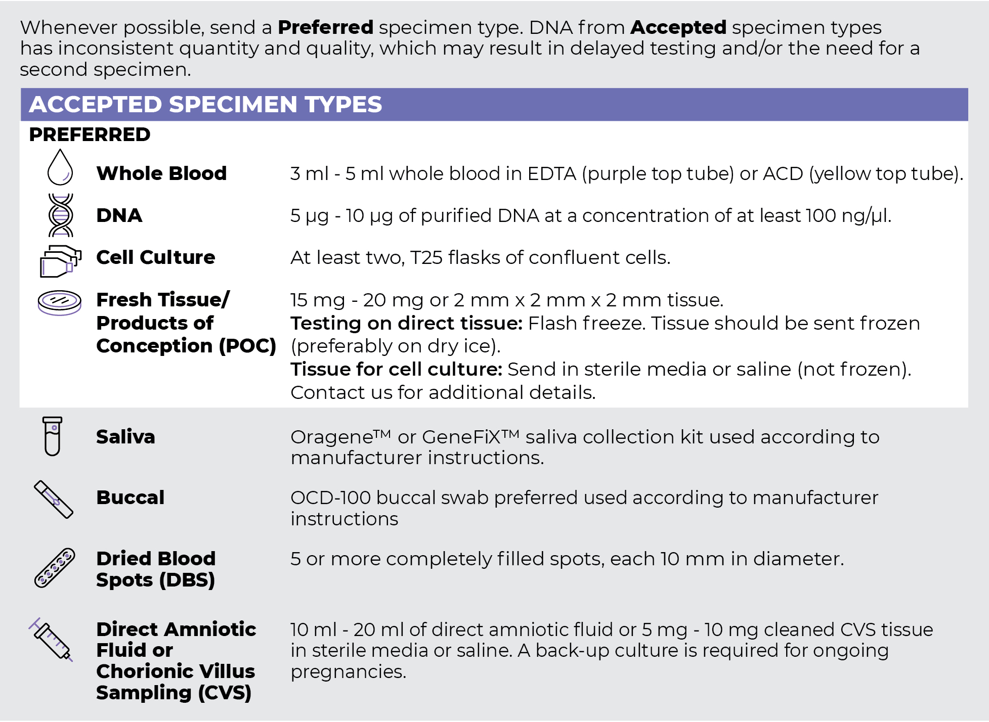
Specimen Types
Specimen Requirements and Shipping Details
ORDER OPTIONS
View Ordering Instructions1) Select Test Type
2) Select Additional Test Options
No Additional Test Options are available for this test.