


C12orf65-Associated Optic Atrophy via the MTRFR/C12orf65 Gene
Summary and Pricing 
Test Method
Exome Sequencing with CNV Detection| Test Code | Test Copy Genes | Test CPT Code | Gene CPT Codes Copy CPT Code | Base Price | |
|---|---|---|---|---|---|
| 11117 | MTRFR | 81479 | 81479,81479 | $990 | Order Options and Pricing |
Pricing Comments
Our favored testing approach is exome based NextGen sequencing with CNV analysis. This will allow cost effective reflexing to PGxome or other exome based tests. However, if full gene Sanger sequencing is desired for STAT turnaround time, insurance, or other reasons, please see link below for Test Code, pricing, and turnaround time information. If the Sanger option is selected, CNV detection may be ordered through Test #600.
An additional 25% charge will be applied to STAT orders. STAT orders are prioritized throughout the testing process.
Click here for costs to reflex to whole PGxome (if original test is on PGxome Sequencing platform).
Click here for costs to reflex to whole PGnome (if original test is on PGnome Sequencing platform).
The Sanger Sequencing method for this test is NY State approved.
For Sanger Sequencing click here.Turnaround Time
3 weeks on average for standard orders or 2 weeks on average for STAT orders.
Please note: Once the testing process begins, an Estimated Report Date (ERD) range will be displayed in the portal. This is the most accurate prediction of when your report will be complete and may differ from the average TAT published on our website. About 85% of our tests will be reported within or before the ERD range. We will notify you of significant delays or holds which will impact the ERD. Learn more about turnaround times here.
Targeted Testing
For ordering sequencing of targeted known variants, go to our Targeted Variants page.
Clinical Features and Genetics
Clinical Features
Optic Atrophy (OA) is the most prevalent inherited optic neuropathy besides Leber’s hereditary optic neuropathy (LHON). Both share a common pathological hallmark, the preferential loss of retinal ganglion cells (RGCs) (Carelli et al. 2009; Yu-Wai-Man et al. 2010). OA is clinically characterized by bilateral reduction in visual acuity that progresses insidiously from early childhood (Yu-Wai-Man et al. 2011). Other symptoms include central or near central scotomas, tritanopia, variable degree of ptosis, central visual field defects and/or ophthalmalgia and optic nerve pallor. The most common OA is inherited in an autosomal dominant (AD) mode (DOA). Phenotype-genotype studies found that 20% of DOA patients develop a more severe phenotype called “DOA plus” (DOA+), which is characterized by extraocular multi-systemic features, including neurosensory hearing loss, or less commonly chronic progressive external ophthalmoplegia, myopathy, peripheral neuropathy, multiple sclerosis-like illness, spastic paraplegia or cataracts (Yu-Wai-Man et al. 2010; Amati-Bonneau et al. 2009). Disease prevalence is estimated at ~1/30,000 in most populations in the world, but in Denmark it can reach to 1/10,000 due to a founder effect (Kjer et al. 1996; Thiselton et al. 2001; Lenaers et al. 2012).Combined oxidative phosphorylation deficiencies (COXPD) are a common cause of mitochondrial disease. MTRFR-associated COXPD type 7 often results in Leigh syndrome (LS) (Heidary et al. 2014). LS is a neurodegenerative disease which is usually evident in the 1st year of life, but can occur later. Clinical symptoms often include motor and/or intellectual developmental delay, respiratory difficulties, nystagmus, opthalmoparesis, optic atrophy, ataxia, and dystonia (Dahl 1998).Clinically and genetically heterogeneous hereditary spastic paraplegia (HSP) is a group of disorders in which primary symptom is insidiously progressive spasticity (rigid muscles) and weakness of the lower limbs. HSP affects 1 in 10,000 people in the Western world (Polo et al. 1993). The Complicated form of the HSP shows additional neurological signs such as amyotrophy, mental retardation, pigmentary retinal degeneration, optic atrophy, extrapyramidal features, cerebellar ataxia, ichthyosis etc. (Harding 1981; Polo et al. 1993). MTRFR-associated HSP is known as SPG55 (Shimazaki et al. 2012).Charcot-Marie Tooth disease (CMT) is a clinically and genetically heterogeneous group of disorders. MTRFR-associated CMT type 6 is characterized by the onset of neuropathy in childhood and optic atrophy in the second decade of life (Tucci et al. 2014).
Genetics
Mutations in MTRFR have been shown to be causative for autosomal recessive COXPD7, SPG55 and CMT6. These disorders all have axonal neuropathy and optic atrophy symptoms in common (Tucci et al. 2014; Dahl 1998; Shimazaki et al. 2012).MTRFR encoded protein belongs to a family of Class I peptide release factors (RFs) that recognize stop codons and catalyze the ribosomal release of the newly synthesized peptide (Duarte et al. 2012). MTRFR is involved in rescuing stalled mitoribosomes during translation and is essential for cell vitality and mitochondrial function (Kogure et al. 2012). So far, very few (~5) causative mutations have been reported in MTRFR (Human Gene Mutation Database).
Clinical Sensitivity - Sequencing with CNV PGxome
Predicting clinical sensitivity for the MTRFR gene is challenging due to genetic heterogeneity of optic atrophy. However, all the reported causative mutations are detectable by this method. Gross deletions or duplications have not been reported in this gene (Human Gene Mutation Database).
Testing Strategy
This test provides full coverage of all coding exons of the MTRFR gene plus 10 bases of flanking noncoding DNA in all available transcripts along with other non-coding regions in which pathogenic variants have been identified at PreventionGenetics or reported elsewhere. We define full coverage as >20X NGS reads or Sanger sequencing. PGnome panels typically provide slightly increased coverage over the PGxome equivalent. PGnome sequencing panels have the added benefit of additional analysis and reporting of deep intronic regions (where applicable).
Dependent on the sequencing backbone selected for this testing, discounted reflex testing to any other similar backbone-based test is available (i.e., PGxome panel to whole PGxome; PGnome panel to whole PGnome).
Indications for Test
Patients with symptoms suggestive of inherited optic neuropathy are candidates. This test may also be considered for the reproductive partners of individuals who carry pathogenic variants in MTRFR.
Patients with symptoms suggestive of inherited optic neuropathy are candidates. This test may also be considered for the reproductive partners of individuals who carry pathogenic variants in MTRFR.
Gene
| Official Gene Symbol | OMIM ID |
|---|---|
| MTRFR | 613541 |
| Inheritance | Abbreviation |
|---|---|
| Autosomal Dominant | AD |
| Autosomal Recessive | AR |
| X-Linked | XL |
| Mitochondrial | MT |
Disease
| Name | Inheritance | OMIM ID |
|---|---|---|
| Combined Oxidative Phosphorylation Deficiency 7 | AR | 613559 |
Citations
- Amati-Bonneau P, Milea D, Bonneau D, Chevrollier A, Ferré M, Guillet V, Gueguen N, Loiseau D, Crescenzo M-AP de, Verny C, Procaccio V, Lenaers G, et al. 2009. OPA1-associated disorders: phenotypes and pathophysiology. Int. J. Biochem. Cell Biol. 41: 1855–1865. PubMed ID: 19389487
- Carelli V, Morgia C La, Valentino ML, Barboni P, Ross-Cisneros FN, Sadun AA. 2009. Retinal ganglion cell neurodegeneration in mitochondrial inherited disorders. Biochimica et Biophysica Acta (BBA) - Bioenergetics 1787: 518–528. PubMed ID: 19268652
- Dahl HH. 1998. Getting to the nucleus of mitochondrial disorders: identification of respiratory chain-enzyme genes causing Leigh syndrome. American journal of human genetics 63: 1594. PubMed ID: 9837811
- Duarte I, Nabuurs SB, Magno R, Huynen M. 2012. Evolution and Diversification of the Organellar Release Factor Family. Molecular Biology and Evolution 29: 3497–3512. PubMed ID: 22688947
- Harding AE. 1981. Hereditary “pure” spastic paraplegia: a clinical and genetic study of 22 families. J Neurol Neurosurg Psychiatry 44: 871–883. PubMed ID: 7310405
- Heidary G, Calderwood L, Cox GF, Robson CD, Teot LA, Mullon J, Anselm I. 2014. Optic atrophy and a Leigh-like syndrome due to mutations in the c12orf65 gene: report of a novel mutation and review of the literature. J Neuroophthalmol 34: 39–43. PubMed ID: 24284555
- Human Gene Mutation Database (Bio-base).
- Kjer B, Eiberg H, Kjer P, Rosenberg T. 1996. Dominant optic atrophy mapped to chromosome 3q region. II. Clinical and epidemiological aspects. Acta Ophthalmol Scand 74: 3–7. PubMed ID: 8689476
- Kogure H, Hikawa Y, Hagihara M, Tochio N, Koshiba S, Inoue Y, Güntert P, Kigawa T, Yokoyama S, Nameki N. 2012. Solution structure and siRNA-mediated knockdown analysis of the mitochondrial disease-related protein C12orf65. Proteins 80: 2629–2642. PubMed ID: 22821833
- Lenaers G, Hamel C, Delettre C, Amati-Bonneau P, Procaccio V, Bonneau D, Reynier P, Milea D. 2012. Dominant optic atrophy. Orphanet J Rare Dis 7: 46–46. PubMed ID: 22776096
- Polo JM, Calleja J, Combarros O, Berciano J. 1993. Hereditary“ pure” spastic paraplegia: a study of nine families. Journal of Neurology, Neurosurgery & Psychiatry 56: 175–181. PubMed ID: 8382269
- Shimazaki H, Takiyama Y, Ishiura H, Sakai C, Matsushima Y, Hatakeyama H, Honda J, Sakoe K, Naoi T, Namekawa M, Fukuda Y, Takahashi Y, et al. 2012. A homozygous mutation of C12orf65 causes spastic paraplegia with optic atrophy and neuropathy (SPG55). J. Med. Genet. 49: 777–784. PubMed ID: 23188110
- Thiselton DL, Alexander C, Morris A, Brooks S, Rosenberg T, Eiberg H, Kjer B, Kjer P, Bhattacharya SS, Votruba M. 2001. A frameshift mutation in exon 28 of the OPA1 gene explains the high prevalence of dominant optic atrophy in the Danish population: evidence for a founder effect. Human genetics 109: 498–502. PubMed ID: 11735024
- Tucci A, Liu Y-T, Preza E, Pitceathly RD, Chalasani A, Plagnol V, Land JM, Trabzuni D, Ryten M, Jaunmuktane Z, others. 2014. Novel C12orf65 mutations in patients with axonal neuropathy and optic atrophy. Journal of Neurology, Neurosurgery & Psychiatry 85: 486–492. PubMed ID: 24198383
- Yu-Wai-Man P, Griffiths PG, Burke A, Sellar PW, Clarke MP, Gnanaraj L, Ah-Kine D, Hudson G, Czermin B, Taylor RW, Horvath R, Chinnery PF. 2010. The Prevalence and Natural History of Dominant Optic Atrophy Due to OPA1 Mutations. Ophthalmology 117: 1538–1546.e1. PubMed ID: 20417570
- Yu-Wai-Man P, Shankar SP, Biousse V, Miller NR, Bean LJH, Coffee B, Hegde M, Newman NJ. 2011. Genetic Screening for OPA1 and OPA3 Mutations in Patients with Suspected Inherited Optic Neuropathies. Ophthalmology 118: 558–563. PubMed ID: 21036400
Ordering/Specimens
Ordering Options
We offer several options when ordering sequencing tests. For more information on these options, see our Ordering Instructions page. To view available options, click on the Order Options button within the test description.
myPrevent - Online Ordering
- The test can be added to your online orders in the Summary and Pricing section.
- Once the test has been added log in to myPrevent to fill out an online requisition form.
- PGnome sequencing panels can be ordered via the myPrevent portal only at this time.
Requisition Form
- A completed requisition form must accompany all specimens.
- Billing information along with specimen and shipping instructions are within the requisition form.
- All testing must be ordered by a qualified healthcare provider.
For Requisition Forms, visit our Forms page
If ordering a Duo or Trio test, the proband and all comparator samples are required to initiate testing. If we do not receive all required samples for the test ordered within 21 days, we will convert the order to the most effective testing strategy with the samples available. Prior authorization and/or billing in place may be impacted by a change in test code.
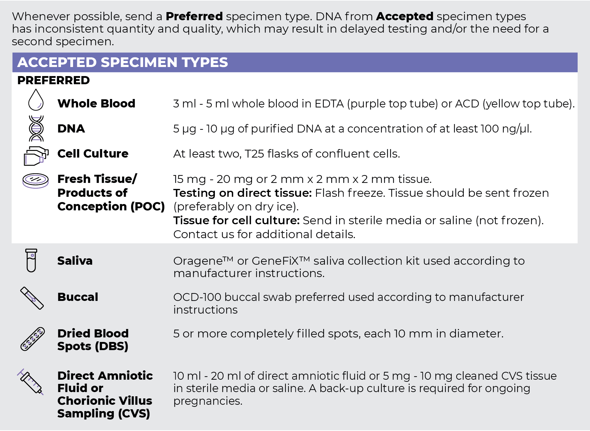
Specimen Types
Specimen Requirements and Shipping Details
PGxome (Exome) Sequencing Panel
PGnome (Genome) Sequencing Panel

ORDER OPTIONS
View Ordering Instructions1) Select Test Type
2) Select Additional Test Options
No Additional Test Options are available for this test.