


TGFBI-Associated Corneal Dystrophies via the TGFBI Gene
Summary and Pricing 
Test Method
Exome Sequencing with CNV Detection| Test Code | Test Copy Genes | Test CPT Code | Gene CPT Codes Copy CPT Code | Base Price | |
|---|---|---|---|---|---|
| 8109 | TGFBI | 81479 | 81479,81479 | $990 | Order Options and Pricing |
Pricing Comments
Our favored testing approach is exome based NextGen sequencing with CNV analysis. This will allow cost effective reflexing to PGxome or other exome based tests. However, if full gene Sanger sequencing is desired for STAT turnaround time, insurance, or other reasons, please see link below for Test Code, pricing, and turnaround time information. If the Sanger option is selected, CNV detection may be ordered through Test #600.
An additional 25% charge will be applied to STAT orders. STAT orders are prioritized throughout the testing process.
Click here for costs to reflex to whole PGxome (if original test is on PGxome Sequencing platform).
Click here for costs to reflex to whole PGnome (if original test is on PGnome Sequencing platform).
The Sanger Sequencing method for this test is NY State approved.
For Sanger Sequencing click here.Turnaround Time
3 weeks on average for standard orders or 2 weeks on average for STAT orders.
Please note: Once the testing process begins, an Estimated Report Date (ERD) range will be displayed in the portal. This is the most accurate prediction of when your report will be complete and may differ from the average TAT published on our website. About 85% of our tests will be reported within or before the ERD range. We will notify you of significant delays or holds which will impact the ERD. Learn more about turnaround times here.
Targeted Testing
For ordering sequencing of targeted known variants, go to our Targeted Variants page.
Clinical Features and Genetics
Clinical Features
Corneal dystrophies (CDs) are rare inherited disorders. Clinically, CDs are characterized as loss of corneal transparency and impaired refraction, which may be caused by a progressive accumulation of deposits (which can be amyloid, hyaline or a combination) on different layers of the cornea. With disease progression, visual acuity gradually decreases and can lead to visual impairment (Correa-Gomez et al. 2007; Klintworth 2009). CDs are non-inflammatory corneal diseases that are classified into three groups based on the sole or predominant anatomical location of the deposits. The three groups are anterior CDs, stromal CDs and posterior CDs. Most CDs exhibit autosomal dominant inheritance with a high degree of penetration. However, CDs present marked inter-and intra-familial variation in clinical expressivity (Klintworth 2009; Munier et al. 2002). CDs are typically evident in first or second decades of life, and manifestations are restricted to the cornea (Zenteno et al. 2006; Klintworth 2009).
Genetics
CDs are genetically heterogeneous (Poulaki and Colby 2008). The Stromal CDs ( defined as all lattice CD types (LCDI, LCDIIIA, LCDI/IIIA and LCDIV), granular Groenouw type I (GCDI), and Avellino (ACD)) and the anterior CDs (defined as Thiel-Behnke dystrophy corneal dystrophy of Bowman layer type II (CDB2) and Reis-Bucklers (anterior CD)) are autosomal dominant and caused by mutations in the TGFBI (previously known as BIGH3, beta ig-h3) gene (Stewart et al. 1999; Takács et al. 2007). TGFBI is the Tissue Growth Factor Beta-Induced gene, which is located on chromosome 5q31. TGFBI encodes a protein called “keratoepithelin”, which is preferentially expressed on the extracellular surface of corneal epithelial cells, and might share β1integrin immunologic properties (Escribano et al. 1994). β1integrin is shown to be important for both the adhesion/migration and proliferation/differentiation of T cells (Maguire 1995). Mutation analyses of TGFBI suggests mutation hot spots at two arginine codons Arg124 and Arg 555 due to CpG dinucleotide transitions (Munier et al. 1997; Korvatska et al. 1998; Munier et al. 2002; Yang et al. 2010). So far, over 60 pathogenic variations in TGFBI (missense, nonsense, small insertions/duplications) have been reported in TGFBI-associated CDs (Human Gene Mutation Database).
Clinical Sensitivity - Sequencing with CNV PGxome
A mutation spectrum analysis identified TGFBI mutations in 80% of corneal dystrophy patients (50/61). Approximately 50% (24/50) of these patients had mutations at Arg124 and Arg 555 amino acids, which indicates the mutation hotspot (Munier et al. 2002). Analytical sensitivity is expected to be high as all the reported TGFBI mutations are detectable by direct sequencing of genomic DNA, and no gross deletions have been reported so far (Human Gene Mutation Database).
Testing Strategy
This test provides full coverage of all coding exons of the TGFBI gene plus 10 bases of flanking noncoding DNA in all available transcripts along with other non-coding regions in which pathogenic variants have been identified at PreventionGenetics or reported elsewhere. We define full coverage as >20X NGS reads or Sanger sequencing. PGnome panels typically provide slightly increased coverage over the PGxome equivalent. PGnome sequencing panels have the added benefit of additional analysis and reporting of deep intronic regions (where applicable).
Dependent on the sequencing backbone selected for this testing, discounted reflex testing to any other similar backbone-based test is available (i.e., PGxome panel to whole PGxome; PGnome panel to whole PGnome).
Indications for Test
All patients with symptoms suggestive of Corneal dystrophies are candidates.
All patients with symptoms suggestive of Corneal dystrophies are candidates.
Gene
| Official Gene Symbol | OMIM ID |
|---|---|
| TGFBI | 601692 |
| Inheritance | Abbreviation |
|---|---|
| Autosomal Dominant | AD |
| Autosomal Recessive | AR |
| X-Linked | XL |
| Mitochondrial | MT |
Diseases
Citations
- Correa-Gomez V, Villalvazo-Cordero L, Zenteno JC. 2007. The TGFBI A546D mutation causes an atypical type of lattice corneal dystrophy. Mol Vis 13: 1695–1700. PubMed ID: 17893671
- Escribano J, Hernando N, Ghosh S, Crabb J, Coca-Prados M. 1994. cDNA from human ocular ciliary epithelium homologous to beta ig-h3 is preferentially expressed as an extracellular protein in the corneal epithelium. J. Cell. Physiol. 160: 511–521. PubMed ID: 8077289
- Human Gene Mutation Database (Bio-base).
- Klintworth GK. 2009. Corneal dystrophies. Orphanet Journal of Rare Diseases 4: 7. PubMed ID: 19236704
- Korvatska E, Munier FL, Djemai A, Wang MX, Frueh B, Chiou A-Y, Uffer S, Ballestrazzi E, Braunstein RE, Forster RK, others. 1998. Mutation hot spots in 5q31-linked corneal dystrophies. The American Journal of Human Genetics 62: 320–324. PubMed ID: 9463327
- Maguire JE, Danahey KM, Burkly LC, Seventer GA Van. 1995. T cell receptor-and beta 1 integrin-mediated signals synergize to induce tyrosine phosphorylation of focal adhesion kinase (pp125FAK) in human T cells. The Journal of experimental medicine 182: 2079–2090. PubMed ID: 7500053
- Munier FL, Frueh BE, Othenin-Girard P, Uffer S, Cousin P, Wang MX, Héon E, Black GC, Blasi MA, Balestrazzi E, others. 2002. BIGH3 mutation spectrum in corneal dystrophies. Investigative ophthalmology & visual science 43: 949–954. PubMed ID: 11923233
- Munier FL, Korvatska E, Djemaï A, Paslier D Le, Zografos L, Pescia G, Schorderet DF. 1997. Kerato-epithelin mutations in four 5q31-linked corneal dystrophies. Nat. Genet. 15: 247–251. PubMed ID: 9054935
- Poulaki V, Colby K. 2008. Genetics of anterior and stromal corneal dystrophies. Semin Ophthalmol 23: 9–17. PubMed ID: 18214787
- Stewart HS, Ridgway AE, Dixon MJ, Bonshek R, Parveen R, Black G. 1999. Heterogeneity in granular corneal dystrophy: identification of three causative mutations in the TGFBI (BIGH3) gene-lessons for corneal amyloidogenesis. Hum. Mutat. 14: 126–132. PubMed ID: 10425035
- Takács L, Losonczy G, Matesz K, Balogh I, Sohajda Z, Tóth K, Fazakas F, Vereb G, Berta A. 2007. TGFBI (BIGH3) gene mutations in Hungary—report of the novel F547S mutation associated with polymorphic corneal amyloidosis. Mol Vis 13: 1976–1983. PubMed ID: 17982422
- Yang J, Han X, Huang D, Yu L, Zhu Y, Tong Y, Zhu B, Li C, Weng M, Ma X. 2010. Analysis of TGFBI gene mutations in Chinese patients with corneal dystrophies and review of the literature. Molecular vision 16: 1186. PubMed ID: 20664689
- Zenteno JC, Ramirez-Miranda A, Santacruz-Valdes C, Suarez-Sanchez R. 2006. Expanding the mutational spectrum in TGFBI-linked corneal dystrophies: identification of a novel and unusual mutation (Val113Ile) in a family with granular dystrophy. Mol Vis 12: 331–335. PubMed ID: 16636649
Ordering/Specimens
Ordering Options
We offer several options when ordering sequencing tests. For more information on these options, see our Ordering Instructions page. To view available options, click on the Order Options button within the test description.
myPrevent - Online Ordering
- The test can be added to your online orders in the Summary and Pricing section.
- Once the test has been added log in to myPrevent to fill out an online requisition form.
- PGnome sequencing panels can be ordered via the myPrevent portal only at this time.
Requisition Form
- A completed requisition form must accompany all specimens.
- Billing information along with specimen and shipping instructions are within the requisition form.
- All testing must be ordered by a qualified healthcare provider.
For Requisition Forms, visit our Forms page
If ordering a Duo or Trio test, the proband and all comparator samples are required to initiate testing. If we do not receive all required samples for the test ordered within 21 days, we will convert the order to the most effective testing strategy with the samples available. Prior authorization and/or billing in place may be impacted by a change in test code.
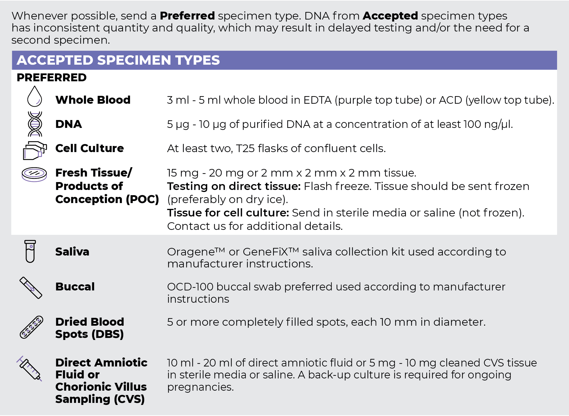
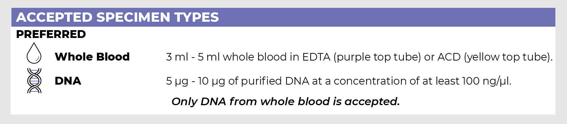
Specimen Types
Specimen Requirements and Shipping Details
PGxome (Exome) Sequencing Panel
PGnome (Genome) Sequencing Panel
ORDER OPTIONS
View Ordering Instructions1) Select Test Type
2) Select Additional Test Options
No Additional Test Options are available for this test.