


GLUT1 Deficiency Syndrome via the SLC2A1 Gene
Summary and Pricing 
Test Method
Exome Sequencing with CNV Detection| Test Code | Test Copy Genes | Test CPT Code | Gene CPT Codes Copy CPT Code | Base Price | |
|---|---|---|---|---|---|
| 3813 | SLC2A1 | 81405 | 81405,81479 | $990 | Order Options and Pricing |
An additional 25% charge will be applied to STAT orders. STAT orders are prioritized throughout the testing process.
Click here for costs to reflex to whole PGxome (if original test is on PGxome Sequencing platform).
Click here for costs to reflex to whole PGnome (if original test is on PGnome Sequencing platform).
Turnaround Time
3 weeks on average for standard orders or 2 weeks on average for STAT orders.
Please note: Once the testing process begins, an Estimated Report Date (ERD) range will be displayed in the portal. This is the most accurate prediction of when your report will be complete and may differ from the average TAT published on our website. About 85% of our tests will be reported within or before the ERD range. We will notify you of significant delays or holds which will impact the ERD. Learn more about turnaround times here.
Targeted Testing
For ordering sequencing of targeted known variants, go to our Targeted Variants page.
Clinical Features and Genetics
Clinical Features
GLUT1 Deficiency Syndrome (GLUT1-DS, also known as De Vivo Syndrome) is a disorder caused by defective glucose transport into the brain. The classical phenotype is characterized by infantile onset seizures which are often refractory, delayed neurological development, acquired microcephaly, and complex movement disorders, which generally consist of ataxia, dystonia and chorea. In approximately 90% of classic GLUT1-DS individuals, seizures begin before two years of age, while they first begin later in life in the remaining 10%. Cognitive impairment is often observed, and may range from mild learning disabilities to severe intellectual disability. The complex movement disorder may be continuous or paroxysmal, and the severity may be influenced by environmental factors, such as hunger, fever, exercise or anxiety (De Giorgis and Veggiotti 2013; Wang et al. 2015).
The clinical definition of GLUT1-DS has been expanded to include non-classical presentations, which includes, but may not be limited to, atypical childhood absence epilepsy, myoclonic epilepsy, intermittent ataxia, choreoathetosis, sleep disturbances, limb dystonia, alternating hemiplegia of childhood with or without epilepsy, paroxysmal exercise-induced dyskinesia and epilepsy (previously known as dystonia 18) and choreoathetosis and spasticity (previously known as dystonia 9) (De Giorgis and Veggiotti 2013; Wang et al. 2015).
Aside from clinical features, certain biomarkers are helpful in diagnosing GLUT1-DS. All individuals with GLUT1-DS have a lower than normal ratio of cerebrospinal fluid (CSF) glucose concentration to blood glucose concentration. Individuals with this syndrome typically have a ratio of less than or equal to ~0.5, while normal individuals have a ratio of 0.6 or greater (De Giorgis and Veggiotti 2013). Additionally, the concentration of CSF lactate is found to be in the low to normal range, and has never been observed to be elevated in a GLUT1-DS individual (De Giorgis and Veggiotti 2013; Wang et al. 2015). Lastly, the majority of individuals with GLUT1-DS have a decreased rate of 3-O-methyl-D-Glucose (3-OMG) uptake in erythrocytes (Yang et al. 2011).
The generally recommended treatment for GLUT1-DS individuals is to be placed on some variation of a ketogenic diet, which is a high-fat, adequate-protein, low-carbohydrate diet. As individuals with GLUT1-DS are unable to transport an adequate amount of glucose into the brain, the ketogenic diet results in a higher than normal level of ketone bodies being generated. These are able to be transported into the brain for use as an alternative fuel source. For the majority of GLUT1-DS patients, such dietary control helps to greatly decrease the frequency of seizures, and often helps decrease the severity of the movement disorder as well (De Giorgis and Veggiotti 2013; Wang et al. 2015).
Pathogenic variants in the SLC2A1 gene have also been reported in two patients with cryohydrocytosis. Clinical findings have included hemolytic anemia upon exposure to cold, hepatosplenomegaly, cataracts, seizures, intellectual disability and movement disorder (Flatt et al. 2011).
Genetics
GLUT1-DS is only known to be caused by defects in the SLC2A1 gene. This disorder is most commonly inherited in an autosomal dominant fashion, although autosomal recessive inheritance has been reported in two unrelated families (Klepper et al. 2009; Rotstein et al. 2010; Wang et al. 2015). Of those with autosomal dominant GLUT1-DS, approximately 90% are found to have a de novo heterozygous pathogenic variant, while approximately 10% inherit a pathogenic variant from a parent. Heterozygous parents tend to have a mild phenotype or be asymptomatic, suggestive of mosaicism. Penetrance of autosomal dominant GLUT1-DS is considered to be complete (De Giorgis and Veggiotti 2013; Wang et al. 2015).
To date, over 160 pathogenic variants have been reported in the SLC2A1 gene, including missense, nonsense, splice site, translation initiation, small insertions, deletions and indels, and multi-exonic and whole gene deletions (Human Gene Mutation Database). The variants are generally spread throughout the gene, although a few mutation “hot spots” have been reported. These include residues p.Arg126, p.Arg333, and the entirety of exon 4 (De Giorgis and Veggiotti 2013; Leen et al. 2010; Wang et al. 2015). A few studies have shown a potential correlation between the type of variant and severity of the disease, with missense variants being most commonly associated with mild to moderate presentation, protein truncating variants being associated primarily with moderate to severe presentations, and exonic or whole gene deletions being associated with severe presentations (Leen et al. 2010; Yang et al. 2011).
The SLC2A1 gene encodes the GLUT1 glucose transporter, which is the fundamental transporter that aids in the facilitated diffusion of glucose across the blood-brain barrier to supply the brain with glucose (De Giorgis and Veggiotti 2013; Wang et al. 2015). The GLUT1 transporter is an integral membrane protein, with twelve transmembrane helices and a pore through which glucose is transported. Pathogenic missense variants have been found to cluster in the regions of the protein involved in substrate binding, pore gating, and lining the transport path (Deng et al. 2014). As glucose is the primary energy source for brain metabolism, defects in the GLUT1 transporter can lead to profound neurological effects (De Giorgis and Veggiotti 2013).
Clinical Sensitivity - Sequencing with CNV PGxome
Clinical sensitivity has been reported to be 40-65% when patients are identified based solely on clinical features or a combination of clinical features, CSF glucose and lactate concentrations and blood glucose concentration (Leen et al. 2010; Yang et al. 2011). Out of 132 patients with GLUT1-DS suspected based solely on clinical features, Leen and colleagues identified pathogenic sequence changes in 57 individuals (Leen et al. 2010). Of those 57 individuals, 51 harbored DNA variants that were detectable via DNA sequencing, suggesting a clinical sensitivity of ~89% in patients with confirmed GLUT1-DS.
Yang and colleagues examined 109 patients for GLUT1-DS (Yang et al. 2011). This group of patients had clinical features consistent with the classical phenotype, as well as expected results for CSF glucose and lactate concentrations and blood glucose concentration. Prior to molecular genetic analysis, the patient samples were further stratified based on the results of the 3-O-methyl-D-Glucose (3-OMG) uptake assay. Of the 109 initial patients, 74 were found to have decreased 3-OMG uptake, while 35 had normal 3-OMG uptake. Of those with decreased 3-OMG uptake, 70 patients were found to harbor pathogenic SLC2A1 variants, of which ~81% were variants detectable via DNA sequencing.
Overall, these results suggest a clinical sensitivity of ~80-90% in individuals with GLUT1-DS who exhibit typical clinical features, laboratory results and decreased 3-OMG uptake assay results. It should be noted Yang et al. (2011) identified one individual with a pathogenic SLC2A1 missense variant that had normal 3-OMG uptake results, so patients with such results should not necessarily be ruled out for molecular genetic testing if other clinical features fit a diagnosis of GLUT1-DS.
While the majority of SLC2A1 variants are expected to be detected via gene sequencing, greater than 10% of reported variants in this gene are exonic or whole-gene deletions, not detectable via sequencing. Out of 57 individuals with pathogenic molecular variations in the SLC2A1 gene, Leen and colleagues identified six with deletions encompassing multiple exons (~11%) (Leen et al. 2010). Similarly, Yang et al. identified five individuals with large intragenic deletions and five with whole-gene deletions in a group of 74 patients with decreased 3-OMG uptake (~14%) (Yang et al. 2011).
Testing Strategy
This test provides full coverage of all coding exons of the SLC2A1 gene plus 10 bases of flanking noncoding DNA in all available transcripts along with other non-coding regions in which pathogenic variants have been identified at PreventionGenetics or reported elsewhere. We define full coverage as >20X NGS reads or Sanger sequencing. PGnome panels typically provide slightly increased coverage over the PGxome equivalent. PGnome sequencing panels have the added benefit of additional analysis and reporting of deep intronic regions (where applicable).
Dependent on the sequencing backbone selected for this testing, discounted reflex testing to any other similar backbone-based test is available (i.e., PGxome panel to whole PGxome; PGnome panel to whole PGnome).
Indications for Test
Patients with clinical symptoms of GLUT1 Deficiency Syndrome are good candidates for this test, particularly if they have been shown to have a low concentration of glucose in their CSF and a low CSF to blood glucose concentration ratio (De Giorgis and Veggiotti 2013). Patients who, in addition, have demonstrated decreased erythrocyte glucose uptake in the 3-O-methyl-D-Glucose (3-OMG) assay are especially good candidates (Yang et al. 2011). Family members of patients who have known SLC2A1 variants are good candidates for this test. We will also sequence the SLC2A1 gene to determine carrier status and to confirm research results.
Patients with clinical symptoms of GLUT1 Deficiency Syndrome are good candidates for this test, particularly if they have been shown to have a low concentration of glucose in their CSF and a low CSF to blood glucose concentration ratio (De Giorgis and Veggiotti 2013). Patients who, in addition, have demonstrated decreased erythrocyte glucose uptake in the 3-O-methyl-D-Glucose (3-OMG) assay are especially good candidates (Yang et al. 2011). Family members of patients who have known SLC2A1 variants are good candidates for this test. We will also sequence the SLC2A1 gene to determine carrier status and to confirm research results.
Gene
| Official Gene Symbol | OMIM ID |
|---|---|
| SLC2A1 | 138140 |
| Inheritance | Abbreviation |
|---|---|
| Autosomal Dominant | AD |
| Autosomal Recessive | AR |
| X-Linked | XL |
| Mitochondrial | MT |
Diseases
| Name | Inheritance | OMIM ID |
|---|---|---|
| Dystonia 9 | AD | 601042 |
| Epilepsy, Idiopathic Generalized, Suscpetibility to, 12 | AD | 614847 |
| Glut1 Deficiency Syndrome 1 | AR, AD | 606777 |
| Glut1 Deficiency Syndrome 2 | AD | 612126 |
Citations
- De Giorgis V, Veggiotti P. 2013. GLUT1 deficiency syndrome 2013: current state of the art. Seizure 22: 803–811. PubMed ID: 23890838
- Deng D, Xu C, Sun P, Wu J, Yan C, Hu M, Yan N. 2014. Crystal structure of the human glucose transporter GLUT1. Nature 510: 121–125. PubMed ID: 24847886
- Flatt JF, Guizouarn H, Burton NM, Borgese F, Tomlinson RJ, Forsyth RJ, Baldwin SA, Levinson BE, Quittet P, Aguilar-Martinez P, Delaunay J, Stewart GW, et al. 2011. Stomatin-deficient cryohydrocytosis results from mutations in SLC2A1: a novel form of GLUT1 deficiency syndrome. Blood 118: 5267–5277. PubMed ID: 21791420
- Human Gene Mutation Database (Bio-base).
- Klepper J, Scheffer H, Elsaid MF, Kamsteeg E-J, Leferink M, Ben-Omran T. 2009. Autosomal recessive inheritance of GLUT1 deficiency syndrome. Neuropediatrics 40: 207–210. PubMed ID: 20221955
- Leen WG, Klepper J, Verbeek MM, Leferink M, Hofste T, Engelen BG van, Wevers RA, Arthur T, Bahi-Buisson N, Ballhausen D, Bekhof J, Bogaert P van, Carrilho I, Chabrol B, Champion MP, Coldwell J, Clayton P, Donner E, Evangeliou A, Ebinger F, Farrell K, Forsyth RJ, de Goede CG, Gross S, Grunewald S, Holthausen H, Jayawant S, Lachlan K, Laugel V, Leppig K, Lim MJ, Mancini G, Marina AD, Martorell L, McMenamin J, Meuwissen ME, Mundy H, Nilsson NO, Panzer A, Poll-The BT, Rauscher C, Rouselle CM, Sandvig I, Scheffner T, Sheridan E, Simpson N, Sykora P, Tomlinson R, Trounce J, Webb D, Weschke B, Scheffer H, Willemsen MA. 2010. Glucose transporter-1 deficiency syndrome: the expanding clinical and genetic spectrum of a treatable disorder. Brain 133: 655–670. PubMed ID: 20129935
- Rotstein M, Engelstad K, Yang H, Wang D, Levy B, Chung WK, Vivo DC De. 2010. Glut1 deficiency: inheritance pattern determined by haploinsufficiency. Ann. Neurol. 68: 955–958. PubMed ID: 20687207
- Wang D, Pascual JM, De Vivo D. 2015. Glucose Transporter Type 1 Deficiency Syndrome. In: Pagon RA, Adam MP, Ardinger HH, et al., editors. GeneReviews™, Seattle (WA): University of Washington, Seattle. PubMed ID: 20301603
- Yang H, Wang D, Engelstad K, Bagay L, Wei Y, Rotstein M, Aggarwal V, Levy B, Ma L, Chung WK, Vivo DC De. 2011. Glut1 deficiency syndrome and erythrocyte glucose uptake assay. Ann. Neurol. 70: 996–1005. PubMed ID: 22190371
Ordering/Specimens
Ordering Options
We offer several options when ordering sequencing tests. For more information on these options, see our Ordering Instructions page. To view available options, click on the Order Options button within the test description.
myPrevent - Online Ordering
- The test can be added to your online orders in the Summary and Pricing section.
- Once the test has been added log in to myPrevent to fill out an online requisition form.
- PGnome sequencing panels can be ordered via the myPrevent portal only at this time.
Requisition Form
- A completed requisition form must accompany all specimens.
- Billing information along with specimen and shipping instructions are within the requisition form.
- All testing must be ordered by a qualified healthcare provider.
For Requisition Forms, visit our Forms page
If ordering a Duo or Trio test, the proband and all comparator samples are required to initiate testing. If we do not receive all required samples for the test ordered within 21 days, we will convert the order to the most effective testing strategy with the samples available. Prior authorization and/or billing in place may be impacted by a change in test code.
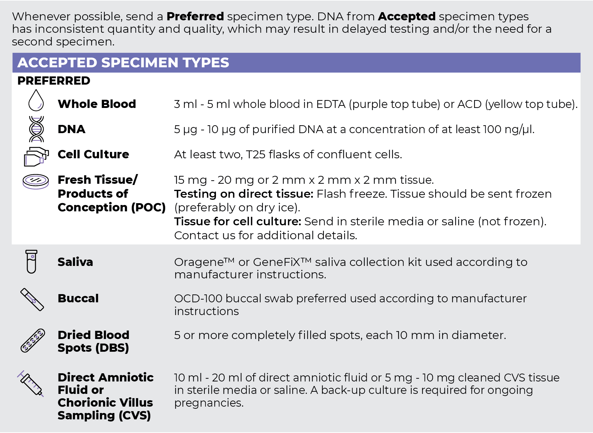
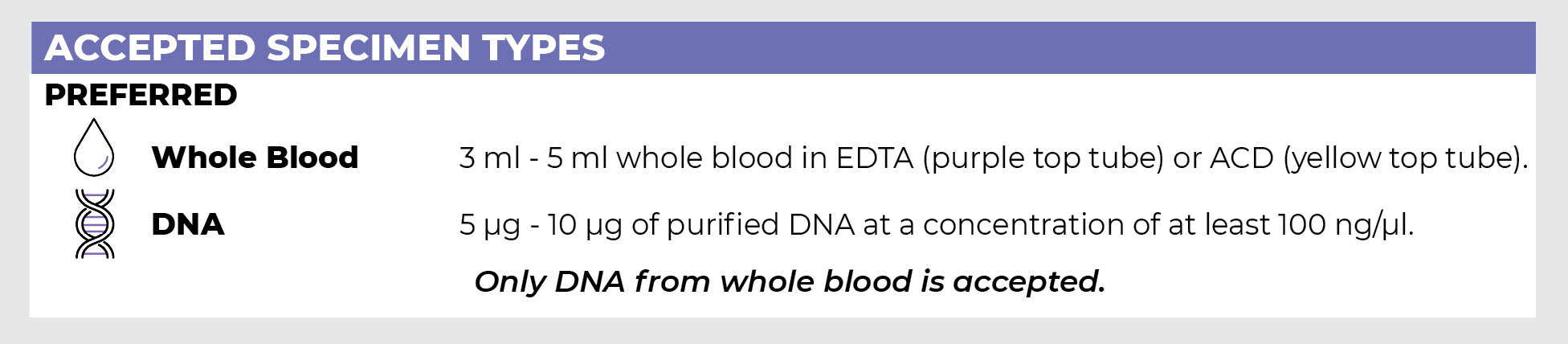
Specimen Types
Specimen Requirements and Shipping Details
PGxome (Exome) Sequencing Panel
PGnome (Genome) Sequencing Panel
ORDER OPTIONS
View Ordering Instructions1) Select Test Type
2) Select Additional Test Options
No Additional Test Options are available for this test.