


Best Vitelliform Macular Dystrophy (BVMD) and Bestrophinopathies via the BEST1 Gene
Summary and Pricing 
Test Method
Exome Sequencing with CNV Detection| Test Code | Test Copy Genes | Test CPT Code | Gene CPT Codes Copy CPT Code | Base Price | |
|---|---|---|---|---|---|
| 8273 | BEST1 | 81406 | 81406,81479 | $990 | Order Options and Pricing |
Pricing Comments
Our favored testing approach is exome based NextGen sequencing with CNV analysis. This will allow cost effective reflexing to PGxome or other exome based tests. However, if full gene Sanger sequencing is desired for STAT turnaround time, insurance, or other reasons, please see link below for Test Code, pricing, and turnaround time information. If the Sanger option is selected, CNV detection may be ordered through Test #600.
An additional 25% charge will be applied to STAT orders. STAT orders are prioritized throughout the testing process.
Click here for costs to reflex to whole PGxome (if original test is on PGxome Sequencing platform).
Click here for costs to reflex to whole PGnome (if original test is on PGnome Sequencing platform).
The Sanger Sequencing method for this test is NY State approved.
For Sanger Sequencing click here.Turnaround Time
3 weeks on average for standard orders or 2 weeks on average for STAT orders.
Please note: Once the testing process begins, an Estimated Report Date (ERD) range will be displayed in the portal. This is the most accurate prediction of when your report will be complete and may differ from the average TAT published on our website. About 85% of our tests will be reported within or before the ERD range. We will notify you of significant delays or holds which will impact the ERD. Learn more about turnaround times here.
Targeted Testing
For ordering sequencing of targeted known variants, go to our Targeted Variants page.
Clinical Features and Genetics
Clinical Features
Visual acuity in humans and other higher primates is mediated by the central (or macular) region of the retina called the fovea, which has the highest density of cone photoreceptor cells (Sun et al. Proc Natl Acad Sci USA 99(6):4008-4013, 2002). Age related-Macular degeneration (AMD) is the most common cause of irreversible vision loss that usually affects older adults due to progressive changes in the retina and/or the underlying retinal pigment epithelium (RPE). Both genetic and environmental factors influence AMD, but juvenile-onset macular degeneration (MD) or Best vitelliform macular dystrophy (BVMD/Best disease, OMIM 153700) is exclusively a genetic disorder (Hartzell et al. Physiology 20:292-302, 2005). Autosomal dominant (AD) BVMD is one of the most common retinal degeneration disorders, with an estimated prevalence of 1.5 per 100 000 individuals in Denmark (Bitner, H. et al. Am J Ophthalmol 154(2):403-412, 2012). BVMD is clinically characterized by large deposits of lipofuscin-like material in the RPE, which forms the characteristic macular lesions resembling the egg yolk ('vitelliform'), a normal electoretinogram (ERG) and an unusual electrooculogram (EOG) light rise and risk of angle-closure glaucoma (Low et al. Mol Vis 17:2272-2282, 2011). The BEST1 gene (Gene/Locus OMIM 607854; previously known as VMD2) has been linked to BVMD. Over 250 causative mutations have been identified in this gene and are associated with clinically distinct ocular phenotypes that are collectively referred as bestrophinopathies (Piñeiro-Gallego et al. Mol Vis 17:1607-1617, 2011). The other clinical phenotypes associated with BEST1 mutations are AD Vitreo-retino-choroidopathy (ADVIRC, OMIM 193220) (Kaufman et al. Arch Ophthalmol 100(2):272-278, 1982), Vitelliform macular dystrophy, adult-onset (AVMD, OMIM 608161)(Do and Ferrucci. Optometry 77(4):156-166, 2006), Autosomal recessive (AR) Bestrophinopathy (ARB, OMIM 611809) (Burgess et al. Am J Hum Genet 82(1):19-31, 2008) and AD/AR Retinitis pigmentosa-50 (RP50, OMIM 613194) (Davidson et al. Am J Hum Genet 85(5):581-592, 2009).
Genetics
While BEST1 mutations typically cause AD inheritance, AR inheritance has also been reported (Bitner, H. et al. Invest Ophthalmol Vis Sci 52(8):5332-5338, 2011). BEST1, located on the long arm (q13) of chromosome 11, encodes Bestrophin, a transmembrane protein that is located in the basolateral portion of the RPE and is responsible for the basolateral Cl- conductance that regulates voltage-dependent Ca2+ channels and generates the light peak. Dysfunction of this transport due to mutations in BEST1 might result in significant accumulation of lipofuscin, ion imbalance in the space around the photoreceptor outer segments and characteristically reduced light peak, which is a pathological hallmark of Best disease. BEST1 is predominantly expressed in RPE and is comprised of 11 exons coding 585 amino acids (aa). Causative mutations in BEST1 are distributed throughout the first 312 aa, and it has been reported that aa 293–311 are especially highly conserved and have functional importance. Mutations in 16 of these 18 aa are linked to Best disease (Hartzell et al. 2005; White et al Hum Mutat 15(4):301-308, 2000). BVMD, AVMD and AMD share some phenotypic features, though AVMD and AMD differ from BVMD by later onset. Along with BEST1 (responsible for 25% of AVMD cases), PRPH2 (periperin/RDS) mutations are linked to AVMD (~36% of cases), indicating genetic heterogeneity of the disorder (Zhuk and Edwards. Mol Vis 12:811-815, 2006). BEST1 pre-mRNA splicing mutations that lead to in-frame deletions are reported in ADVIRC (Yardley et al Invest Ophthalmol Vis Sci 45(10):3683-3689, 2004), whereas missense mutations were associated with AD/AR Retinitis Pigmentosa (Davidson et al. 2009) . The vast majority of identified BEST1 mutations (90/93) in patients with classical Best disease are missense mutations or small in-frame deletions (MacDonald, I.M. and Lee, T., GeneReviews, 2009 ; Bitner, H. et al. Am J Ophthalmol 154(2):403-412, 2012). There are a few reports of de novo mutations in BVMD patients whose parents were phenotypically and genetically unaffected (Apushkin et al. Arch Ophthalmol 124(6):887-889, 2006; Palomba et al. Am J Ophthalmol 129(2):260-262, 2000)
Clinical Sensitivity - Sequencing with CNV PGxome
Krämer et al detected 23 unique BEST1 mutations in 34 of 41 BVMD affected patients. Out of 25 probands who had positive family history, 96% (24/25) had BEST1 mutations, whereas with unknown family history the detection rate was significantly reduced to 69% (11/16) (Krämer et al. Eur J Hum Genet 8(4):286-292, 2000). Marchant et al also reports high detection rate for BEST1 mutations in BVMD patients (at least one mutation in each patient) (Marchant et al. J Med Genet 44(3):e70).
Thus far, no gross insertions/deletions have been identified in BEST1 (Human Gene Mutation Database).
Testing Strategy
This test provides full coverage of all coding exons of the BEST1 gene plus 10 bases of flanking noncoding DNA in all available transcripts along with other non-coding regions in which pathogenic variants have been identified at PreventionGenetics or reported elsewhere. We define full coverage as >20X NGS reads or Sanger sequencing. PGnome panels typically provide slightly increased coverage over the PGxome equivalent. PGnome sequencing panels have the added benefit of additional analysis and reporting of deep intronic regions (where applicable).
Dependent on the sequencing backbone selected for this testing, discounted reflex testing to any other similar backbone-based test is available (i.e., PGxome panel to whole PGxome; PGnome panel to whole PGnome).
Indications for Test
Ideal BEST1 test candidates are BVMD, AVMD, ADVIRC, ARB and AD/AR RP patients.
Ideal BEST1 test candidates are BVMD, AVMD, ADVIRC, ARB and AD/AR RP patients.
Gene
| Official Gene Symbol | OMIM ID |
|---|---|
| BEST1 | 607854 |
| Inheritance | Abbreviation |
|---|---|
| Autosomal Dominant | AD |
| Autosomal Recessive | AR |
| X-Linked | XL |
| Mitochondrial | MT |
Diseases
Related Test
| Name |
|---|
| Retinitis Pigmentosa Panel |
Citations
- Apushkin, M.A. et al. (2006). "Novel de novo mutation in a patient with Best macular dystrophy." Arch Ophthalmol 124(6):887-889. PubMed ID: 16769844
- Bitner, H. et al. (2011). “A homozygous frameshift mutation in BEST1 causes the classical form of Best disease in an autosomal recessive mode.” Invest Ophthalmol Vis Sci 52(8):5332-5338. PubMed ID: 21467170
- Bitner, H. et al. (2012). “Frequency, genotype, and clinical spectrum of best vitelliform macular dystrophy: data from a national center in Denmark.” Am J Ophthalmol 154(2):403-412. PubMed ID: 22633354
- Burgess, R. (2008). “Biallelic mutation of BEST1 causes a distinct retinopathy in humans.” Am J Hum Genet 82(1):19-31. PubMed ID: 18179881
- Davidson, A.E. et al. (2009). “Missense mutations in a retinal pigment epithelium protein, bestrophin-1, cause retinitis pigmentosa.” Am J Hum Genet 85(5):581-592. PubMed ID: 19853238
- Do, P and Ferrucci, S. (2006). "Adult-onset foveomacular vitelliform dystrophy." Optometry 77(4):156-166. PubMed ID: 16567277
- Hartzell, C. et al. (2005). “Looking chloride channels straight in the eye: bestrophins, lipofuscinosis, and retinal degeneration.” Physiology (Bethesda) 20:292-302. PubMed ID: 16174869
- Human Gene Mutation Database (Bio-base).
- Kaufman, S.J. et al. (1982). “Autosomal dominant vitreoretinochoroidopathy.” Arch Ophthalmol 100(2):272-278. PubMed ID: 7065944
- Krämer, F. et al. (2000). “Mutations in the VMD2 gene are associated with juvenile-onset vitelliform macular dystrophy (Best disease) and adult vitelliform macular dystrophy but not age-related macular degeneration.” Eur J Hum Genet 8(4):286-292. PubMed ID: 10854112
- Low, S. et al. (2011). "Autosomal dominant Best disease with an unusual electrooculographic light rise and risk of angle-closure glaucoma: a clinical and molecular genetic study." Mol Vis 17:2272-2282. PubMed ID: 21921978
- MacDonald, I.M. and Lee, T. (2009). "Best Vitelliform Macular Dystrophy." GeneReviews. PubMed ID: 20301346
- Marchant, D. et al. (2007). “New VMD2 gene mutations identified in patients affected by Best vitelliform macular dystrophy.” J Med Genet 44(3):e70. PubMed ID: 17287362
- Palomba, G. et al. (2000). “A novel spontaneous missense mutation in VMD2 gene is a cause of a best macular dystrophy sporadic case.” Am J Ophthalmol 129(2):260-262. PubMed ID: 10682987
- Piñeiro-Gallego, T. et al. (2011). “Clinical evaluation of two consanguineous families with homozygous mutations in BEST1.” Mol Vis 17:1607-1617. PubMed ID: 21738390
- Sun, H. et al. (2002). "The vitelliform macular dystrophy protein defines a new family of chloride channels." Proc Natl Acad Sci USA 99(6):4008-4013. PubMed ID: 11904445
- White, K. et al. (2000). “VMD2 mutations in vitelliform macular dystrophy (Best disease) and other maculopathies.” Hum Mutat 15(4):301-308. PubMed ID: 10737974
- Yardley, J. et al. (2004). “Mutations of VMD2 splicing regulators cause nanophthalmos and autosomal dominant vitreoretinochoroidopathy (ADVIRC).” Invest Ophthalmol Vis Sci 45(10):3683-3689. PubMed ID: 15452077
- Zhuk and Edwards. (2006). "Peripherin/RDS and VMD2 mutations in macular dystrophies with adult-onset vitelliform lesion." Mol Vis 12:811-815. PubMed ID: 16885924
Ordering/Specimens
Ordering Options
We offer several options when ordering sequencing tests. For more information on these options, see our Ordering Instructions page. To view available options, click on the Order Options button within the test description.
myPrevent - Online Ordering
- The test can be added to your online orders in the Summary and Pricing section.
- Once the test has been added log in to myPrevent to fill out an online requisition form.
- PGnome sequencing panels can be ordered via the myPrevent portal only at this time.
Requisition Form
- A completed requisition form must accompany all specimens.
- Billing information along with specimen and shipping instructions are within the requisition form.
- All testing must be ordered by a qualified healthcare provider.
For Requisition Forms, visit our Forms page
If ordering a Duo or Trio test, the proband and all comparator samples are required to initiate testing. If we do not receive all required samples for the test ordered within 21 days, we will convert the order to the most effective testing strategy with the samples available. Prior authorization and/or billing in place may be impacted by a change in test code.
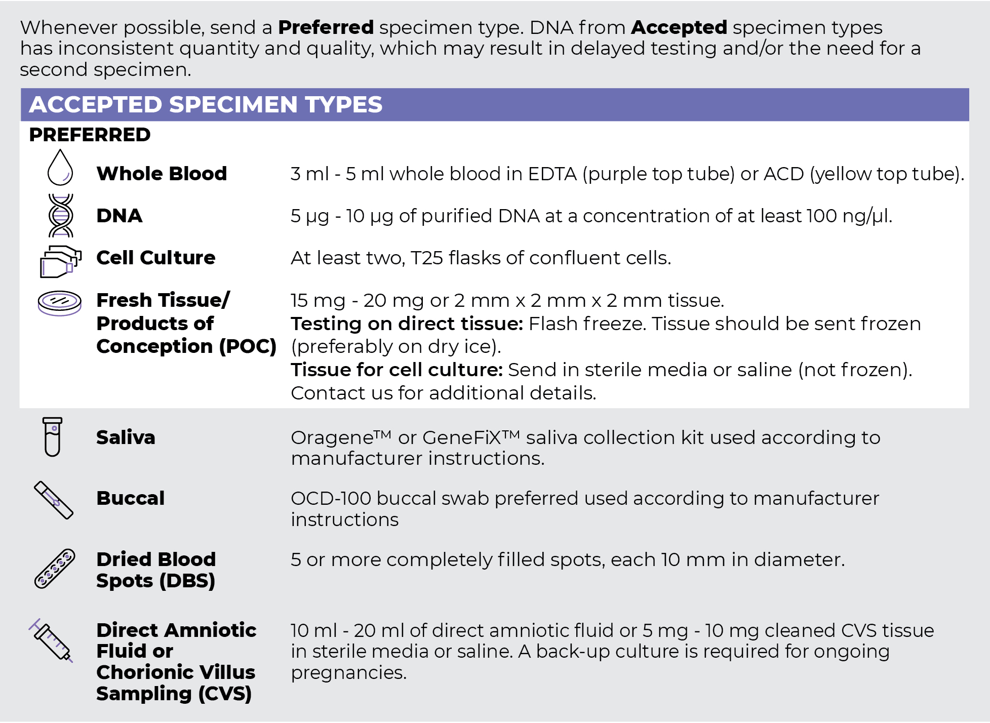
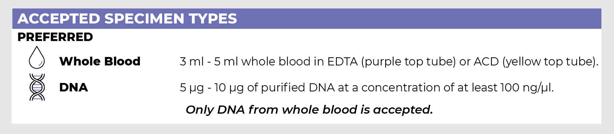
Specimen Types
Specimen Requirements and Shipping Details
PGxome (Exome) Sequencing Panel
PGnome (Genome) Sequencing Panel
ORDER OPTIONS
View Ordering Instructions1) Select Test Type
2) Select Additional Test Options
No Additional Test Options are available for this test.