


Homocystinuria Panel
Summary and Pricing 
Test Method
Exome Sequencing with CNV Detection| Test Code | Test Copy Genes | Panel CPT Code | Gene CPT Codes Copy CPT Code | Base Price | |
|---|---|---|---|---|---|
| 10201 | Genes x (5) | 81479 | 81406(x1), 81479(x9) | $990 | Order Options and Pricing |
Pricing Comments
We are happy to accommodate requests for testing single genes in this panel or a subset of these genes. The price will remain the list price. If desired, free reflex testing to remaining genes on panel is available. Alternatively, a single gene or subset of genes can also be ordered via our Custom Panel tool.
An additional 25% charge will be applied to STAT orders. STAT orders are prioritized throughout the testing process.
Click here for costs to reflex to whole PGxome (if original test is on PGxome Sequencing platform).
Click here for costs to reflex to whole PGnome (if original test is on PGnome Sequencing platform).
Turnaround Time
3 weeks on average for standard orders or 2 weeks on average for STAT orders.
Please note: Once the testing process begins, an Estimated Report Date (ERD) range will be displayed in the portal. This is the most accurate prediction of when your report will be complete and may differ from the average TAT published on our website. About 85% of our tests will be reported within or before the ERD range. We will notify you of significant delays or holds which will impact the ERD. Learn more about turnaround times here.
Targeted Testing
For ordering sequencing of targeted known variants, go to our Targeted Variants page.
Clinical Features and Genetics
Clinical Features
Homocystinuria with hypomethioninemia is a genetic disorder caused by inherited inborn errors in the cobalamin, homocysteine or folate metabolic pathways. Clinical symptoms can be severe, and onset is typically during childhood or infancy, although late-onset cases have been reported. Clinically, patients present with megaloblastic anemia, feeding difficulties, lethargy, hypotonia, cerebral atrophy and developmental delay, ectopia lentis and/or severe myopia, skeletal abnormalities which can include thinning and lengthening of the long bones as well as osteoporosis, and vascular disease, including potentially fatal thromboembolisms. Some patients also exhibit ataxia, neonatal seizures and blindness. Biochemically, patients exhibit homocystinuria, hyperhomocystinemia and hypomethioninemia or hypermethioninemia without methylmalonic aciduria. Affected patients may be identified via newborn screening, although this depends on the methods used by the screening laboratory and, for disorders associated with hypomethioninemia, their ability to detect low levels of methionine. Therefore, complementation analysis and/or molecular genetic testing should still be considered for symptomatic individuals, even if newborn screening results are reported to be normal (Kraus et al. 1999; Carrillo-Carrasco et al. 2013; Picker and Levy 2014; Mudd et al. 2014; Watkins and Rosenblatt 2014).
Four genetic causes of homocystinuria with hypomethionimemia have been identified: cblD variant 1, cblE and cblG types, and homocystinuria due to MTHFR enzyme deficiency. The cblG type of homocystinuria is caused by pathogenic variants in the MTR gene, while the cblE type is caused by pathogenic variants in the MTRR gene. The cblE and cblG disorders are clinically indistinguishable, although the specific diagnosis can be confirmed by enzymatic or cellular complementation assays, as well as molecular genetic studies. The cblE and cblG types of cobalamin metabolic deficiencies can be difficult to distinguish from the cblD variant 1 cobalamin disorder, which is caused by pathogenic variants in the MMADHC gene. Depending on the type and location of the variants found in the MMADHC gene, patients can present with classic cblD type (combined homocystinuria and methylmalonic aciduria), cblD variant 1 (isolated homocystinuria) or cblD variant 2 (isolated methylmalonic aciduria). In addition to symptoms mentioned above, cblD variant 1 patients have also been reported to present with a marfanoid appearance, venous thrombosis, and/or hydrocephalus (Carrillo-Carrasco et al. 2013; Watkins and Rosenblatt 2014). Lastly, pathogenic variants in the MTHFR gene can also cause homocystinuria with low to normal levels of methionine in the plasma (Watkins and Rosenblatt 2014; Froese et al. 2016). It should be noted that MTHFR deficient patients typically do not present with megaloblastic anemia (Watkins and Rosenblatt 2014).
Homocystinuria can also be caused by defects in the CBS (cystathionine beta-synthase) gene. CBS deficiency can typically be biochemically distinguished from the cblD variant 1, cblE and cblG disorders and MTHFR deficiency because hypermethioninemia is observed rather than hypomethioninemia (Mudd et al. 2014). Defects in the CBS enzyme are the most commonly observed cause of homocystinuria without associated methylmalonic aciduria (Picker and Levy 2014; Mudd et al. 2014). Biochemically, patients with this disorder are found to have greatly increased concentrations of plasma and urine homocysteine, total homocysteine, and marked hypermethioninemia (Picker and Levy 2014). Age of onset and expressivity of clinical features displayed by CBS deficienct patients varies widely, even within sibships, and can range from affected newborns to adults who were previously asymptomatic first presenting with a thromboembolytic event (Carrillo-Carrasco et al. 2013; Picker and Levy 2014; Mudd et al. 2014).
For additional information, please see the individual test descriptions for the MTR, MTRR, MMADHC, MTHFR and CBS genes.
Genetics
The cblD variant 1, cblE and cblG types of homocystinuria, as well as MTHFR deficiency and classical CBS deficiency homocystinuria, are all autosomal recessive disorders.
The cblD variant 1 type is caused by variants in the MMADHC gene, and the type and location of the variant(s) affects whether the individual presents with classic cblD, cblD variant 1 or cblD variant 2. The exact function of the MMADHC protein is not known, although it is thought to play a role in the generation or transport of both Adenosylcobalamin (AdoCbl; required by the Methylmalonyl CoA Mutase enzyme) and Methylcobalamin (MeCbl; required by the Methionine Synthase enzyme). To date, fewer than 20 pathogenic variants have been reported in the MMADHC gene. They are a mix of missense, nonsense, small deletion and small insertion variants (Human Gene Mutation Database). The cblG type of homocystinuria is caused by pathogenic variants in the MTR gene, which encodes the Methionine Synthase enzyme. The cblE type is caused by pathogenic variants in the MTRR gene, which encodes the Methionine Synthase Reductase enzyme which is responsible for ensuring the continued function of Methionine Synthase. The MeCbl-dependent Methionine Synthase enzyme is responsible for the conversion of homocysteine to methionine. Over 20 causative variants have been reported in both MTR and MTRR. The majority of reported pathogenic variants are missense and small deletions that lead to premature protein termination, although splice variants, small insertions and indels, and small and gross insertions have also been reported (Human Gene Mutation Database).
MTHFR deficiency and classical CBS deficiency homocystinuria are more commonly reported, with over 100 MTHFR and nearly 200 CBS causative variants documented in the literature (Human Gene Mutation Database). The majority of variants reported in both genes are missense, although protein truncating variants, and in CBS, large deletions, have been reported as well (Human Gene Mutation Database).
Please see individual test descriptions for additional information on the molecular biology of each gene.
Clinical Sensitivity - Sequencing with CNV PGxome
Although the sensitivity of this test panel is currently unknown, most variants reported for the genes in this panel are of the type which can be detected using direct sequencing methods and thus analytical sensitivity is expected to be high. Based on collective totals of reported patient data, the detection rates for pathogenic variants in the MMADHC, MTR and MTRR genes in this panel are approximately as follows: In the MTRR gene, pathogenic variants were detected in 34 of 36 studied alleles, for an overall detection rate of ~94% (Wilson et al. 1999; Zavadáková et al. 2005); In the MTR gene, pathogenic variants were detected in 36 of 42 studied alleles, for an overall detection rate of ~86% (Watkins et al. 2002); In the MMADHC gene, pathogenic variants were detected in all 20 studied alleles, for an overall detection rate of 100% (Coelho et al. 2008; Miousse et al. 2009).
The clinical sensitivity of MTHFR sequencing is expected to be high as to date, nearly all reported patients have had two pathogenic variants detectable via direct MTHFR sequencing (Goyette et al. 1995; Kluijtmans et al. 1998; Sibani et al. 2000; Sibani et al. 2003; Urreizti et al. 2010; Burda et al. 2015). In these studies, a total of 108 patients were reported with 211 alleles carrying a pathogenic variant, suggesting a clinical sensitivity of ~98%.
Lastly, the sensitivity of this CBS sequencing is also expected to be quite high, as most patients reported to date have been found to have two CBS variants detectable via direct sequencing. The majority of studies with larger patient cohorts have reported pathogenic variant detection via direct sequencing in ~95-98% of patient alleles (Gaustadnes et al. 2002; Kruger et al. 2003; Cozar et al. 2011; Karaca et al. 2014).
To date, no gross deletions or insertions have been reported in the MMADHC, MTHFR, MTR or MTRR genes (Human Gene Mutation Database). Therefore, the sensitivity of duplication/deletion testing for these rare disorders, although not precisely known, is low. Large deletions in the CBS gene have been reported, but appear to be a rare cause of disease (Human Gene Mutation Database).
Testing Strategy
This test is performed using Next-Gen sequencing with additional Sanger sequencing as necessary.
This panel provides 100% coverage of all coding exons of the genes plus 10 bases of flanking noncoding DNA in all available transcripts along with other non-coding regions in which pathogenic variants have been identified at PreventionGenetics or reported elsewhere. We define coverage as ≥20X NGS reads or Sanger sequencing. PGnome panels typically provide slightly increased coverage over the PGxome equivalent. PGnome sequencing panels have the added benefit of additional analysis and reporting of deep intronic regions (where applicable).
Dependent on the sequencing backbone selected for this testing, discounted reflex testing to any other similar backbone-based test is available (i.e., PGxome panel to whole PGxome; PGnome panel to whole PGnome).
Please note that as recommended by the ACMG, ACOG and AHA, we do not offer testing specifically for the MTHFR common polymorphisms c.665C>T and c.1286A>C (also known as C677T and A1298C) due to the limited clinical utility of such testing (Hickey et al. 2013; Levin and Varga 2016).
Indications for Test
Individuals with a positive newborn screening result for homocystinuria are good candidates for this test. Additionally, individuals that exhibit biochemical and clinical symptoms of the cblD variant 1, cblE or cblG disorders, CBS or MTHFR deficiency, are good candidates.
Individuals with a positive newborn screening result for homocystinuria are good candidates for this test. Additionally, individuals that exhibit biochemical and clinical symptoms of the cblD variant 1, cblE or cblG disorders, CBS or MTHFR deficiency, are good candidates.
Genes
| Official Gene Symbol | OMIM ID |
|---|---|
| CBS | 613381 |
| MMADHC | 611935 |
| MTHFR | 607093 |
| MTR | 156570 |
| MTRR | 602568 |
| Inheritance | Abbreviation |
|---|---|
| Autosomal Dominant | AD |
| Autosomal Recessive | AR |
| X-Linked | XL |
| Mitochondrial | MT |
Diseases
Related Test
| Name |
|---|
| PGxome® |
Citations
- Burda P. et al. 2015. Human Mutation. 36: 611-21. PubMed ID: 25736335
- Carrillo-Carrasco N. et al. 2013. Disorders of Intracellular Cobalamin Metabolism. In: Pagon RA, Adam MP, Ardinger HH, Bird TD, Dolan CR, Fong C-T, Smith RJ, and Stephens K, editors. GeneReviews(®), Seattle (WA): University of Washington, Seattle. PubMed ID: 20301503
- Coelho et al. 2008. PubMed ID: 18385497
- Cozar M. et al. 2011. Human Mutation. 32: 835-42. PubMed ID: 21520339
- Froese D.S. et al. 2016. Human Mutation. 37: 427-38. PubMed ID: 26872964
- Gaustadnes M. et al. 2002. Human Mutation. 20: 117-26. PubMed ID: 12124992
- Goyette P. et al. 1995. American Journal of Human Genetics. 56: 1052-9. PubMed ID: 7726158
- Hickey S.E. et al. 2013. Genetics in Medicine. 15: 153-6. PubMed ID: 23288205
- Human Gene Mutation Database (Bio-base).
- Karaca M. et al. 2014. Gene. 534: 197-203. PubMed ID: 24211323
- Kluijtmans L.A. et al. 1998. European Journal of Human Genetics. 6: 257-65. PubMed ID: 9781030
- Kraus J.P. et al. 1999. Human Mutation. 13: 362-75. PubMed ID: 10338090
- Kruger W.D. et al. 2003. Human Mutation. 22: 434-41. PubMed ID: 14635102
- Levin B.L., Varga E. 2016. Journal of Genetic Counseling. 25: 901-11. PubMed ID: 27130656
- Miousse et al. 2009. PubMed ID: 19058814
- Mudd et al. 2014. Disorders of Transsulfuration. In: Valle D, Beaudet AL, Vogelstein B, et al., editors.New York, NY: McGraw-Hill. OMMBID.
- Picker J.D. and Levy H.L. 2014. Homocystinuria Caused by Cystathionine Beta-Synthase Deficiency. In: Pagon RA, Adam MP, Ardinger HH, Bird TD, Dolan CR, Fong C-T, Smith RJ, and Stephens K, editors. GeneReviews(®), Seattle (WA): University of Washington, Seattle. PubMed ID: 20301697
- Sibani S. et al. 2000. Human Mutation. 15: 280-7. PubMed ID: 10679944
- Sibani S. et al. 2003. Human Mutation. 21: 509-20. PubMed ID: 12673793
- Urreizti R. et al. 2010. Clinical Genetics. 78: 441-8. PubMed ID: 20236116
- Watkins and Rosenblatt. 2014. Inherited Disorders of Folate and Cobalamin Transport and Metabolism. In: Valle D, Beaudet A.L., Vogelstein B, et al., editors. New York, NY: McGraw-Hill. OMMBID.
- Watkins D. et al. 2002. American Journal of Human Genetics. 71: 143-53. PubMed ID: 12068375
- Wilson A. et al. 1999. Human Molecular Genetics. 8: 2009-16. PubMed ID: 10484769
- Zavadáková P. et al. 2005. Human Mutation. 25: 239-47. PubMed ID: 15714522
Ordering/Specimens
Ordering Options
We offer several options when ordering sequencing tests. For more information on these options, see our Ordering Instructions page. To view available options, click on the Order Options button within the test description.
myPrevent - Online Ordering
- The test can be added to your online orders in the Summary and Pricing section.
- Once the test has been added log in to myPrevent to fill out an online requisition form.
- PGnome sequencing panels can be ordered via the myPrevent portal only at this time.
Requisition Form
- A completed requisition form must accompany all specimens.
- Billing information along with specimen and shipping instructions are within the requisition form.
- All testing must be ordered by a qualified healthcare provider.
For Requisition Forms, visit our Forms page
If ordering a Duo or Trio test, the proband and all comparator samples are required to initiate testing. If we do not receive all required samples for the test ordered within 21 days, we will convert the order to the most effective testing strategy with the samples available. Prior authorization and/or billing in place may be impacted by a change in test code.
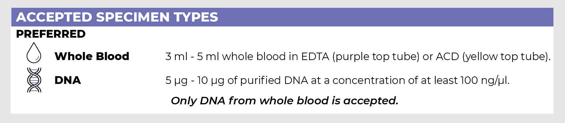
Specimen Types
Specimen Requirements and Shipping Details
PGxome (Exome) Sequencing Panel
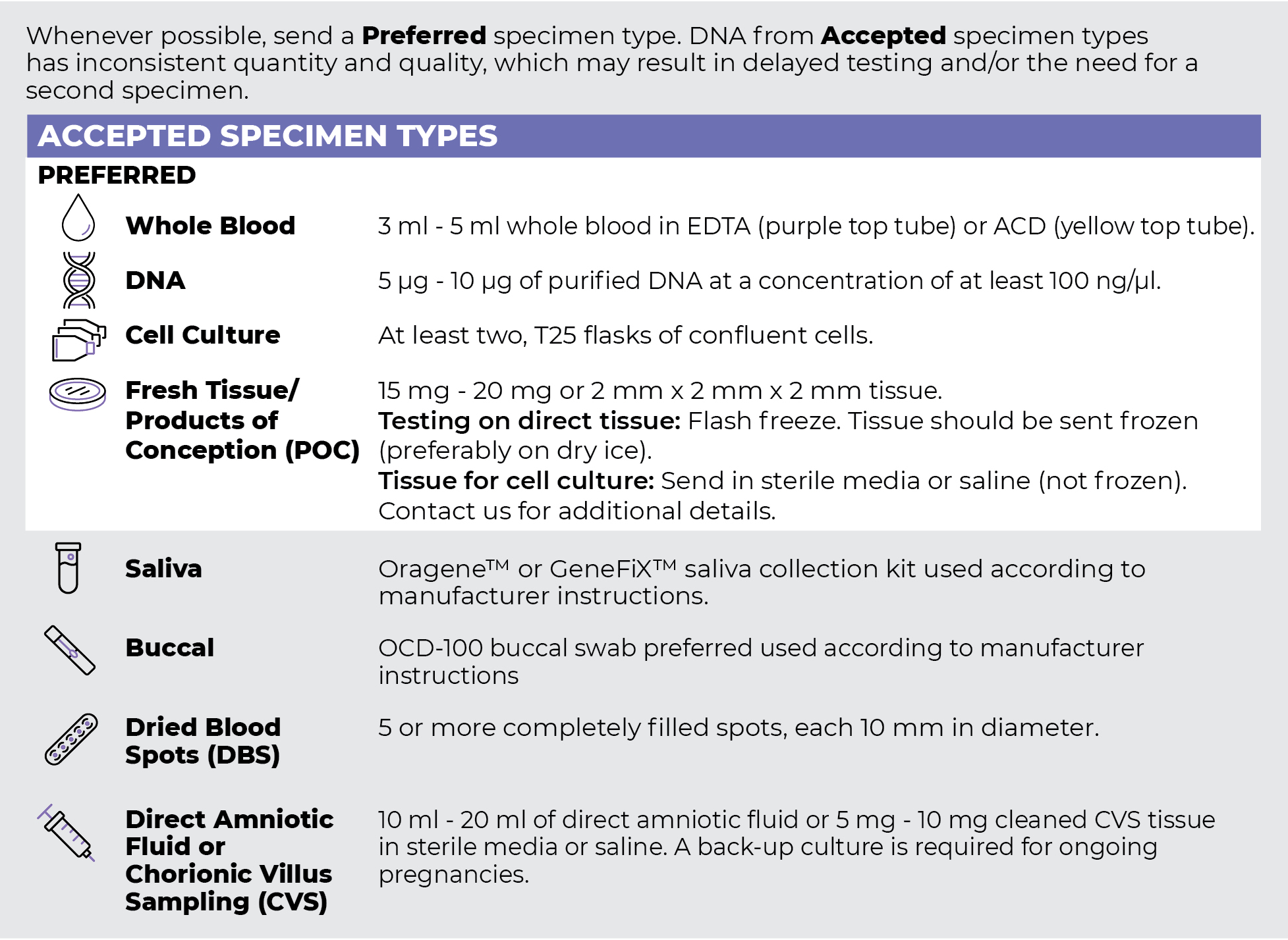
PGnome (Genome) Sequencing Panel
ORDER OPTIONS
View Ordering Instructions1) Select Test Type
2) Select Additional Test Options
No Additional Test Options are available for this test.